How does fatigue develop during aerobic exercise?
Please subscribe to my newsletter to read the future articles in this series!
Fatigue is a temporary reduction in exercise performance as a result of previous exercise. We can analyze the development of fatigue over the course of a single bout of exercise. Recently, I explained how fatigue develops over a strength training set. In this article, explain exactly how fatigue develops over the course of a bout of aerobic exercise.
What mechanisms of fatigue develop during a bout of aerobic exercise?
Introduction
Fatigue is a temporary and reversible reduction in exercise performance due that is usually caused by preceding exercise. During exercise, fatigue can arise through several different fatigue mechanisms, including mechanisms inside the central nervous system (CNS), which lead to reductions in the level of central motor command that reaches the muscle (thereby reducing motor unit recruitment and motor unit firing rates), and mechanisms inside the muscle itself, which lead to reductions in either muscle fiber force or muscle fiber shortening velocity. To understand how fatigue develops over the course of a bout of aerobic exercise, we need to understand exactly how each of these mechanisms takes effect.
The unique features of aerobic exercise
Aerobic exercise differs from strength training in two key respects. Firstly, it involves lower whole muscle forces, and secondly it involves a much longer duration in which muscles are active. These two key features cause fatigue to develop in very different ways. The fact that whole muscle forces are lower is important because metabolite accumulation cannot occur inside a muscle unless whole muscle forces exceed a certain threshold (which is usually 25–30% of maximum isometric force). Therefore, metabolite-related fatigue does not occur during aerobic exercise. For this reason, the only peripheral fatigue mechanisms are those that involve calcium ion accumulation. And the fact that muscles are activated during aerobic exercise for longer than during a strength training set means that the depletion of glycogen stored inside muscle fibers can have a fatiguing effect.
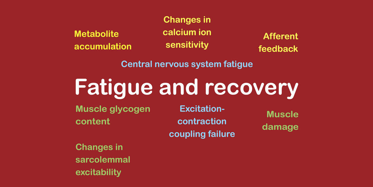
Ultimately, therefore, we can categorize the various fatigue mechanisms that are working during a bout of aerobic exercise into four groups, as follows: [1] glycogen depletion, [2] calcium ion accumulation, [3] spinal CNS fatigue, and [4] supraspinal CNS fatigue. Let’s now take a look at each of these fatigue mechanisms in more detail.
#1. Glycogen depletion
INTRODUCTION
In general, fatigue mechanisms work by reducing the ability of muscle fibers to complete the necessary steps that lead to force production. For example, supraspinal CNS fatigue stops the motor cortex from creating a sufficiently large central motor command such that the high-threshold motor units can be recruited. Similarly, spinal CNS fatigue reduces the size of the central motor command that is transmitted to the neuromuscular junction, stopping certain motor units from being recruited or reducing the motor unit firing rates of the activated muscle fibers. Calcium ion-related fatigue mechanisms stop the electrical signal that is propagated along activated muscle fibers from turning into a chemical signal that triggers crossbridge formation. And finally, metabolite-related fatigue either slows or stops the crossbridge cycle from occurring by interfering directly with the binding of myosin heads to the actin myofilaments. All of these fatigue mechanisms are therefore impairments in the normal process that leads to muscle fibers producing force. Contrary to popular belief, in none of these fatigue mechanisms does a lack of fuel lead to a reduction in force production or power output. Thus, the idea that fatigue typically involves running out of energy is greatly mistaken. Nevertheless, when we consider endurance exercise, a reduction in the availability of glycogen can cause fatigue to occur.
GLYCOGEN DEPLETION
During sustained, intense aerobic exercise, muscle and liver glycogen levels are reduced as the glycogen is broken down to provide fuel for the muscle fiber. The muscle fiber needs fuel to power the crossbridge cycle as well as to power the excitation-contraction coupling process. Thus, glycogen stores are located in several key places throughout muscle fibers, including inside the myofibrils (intramyofibrillar), between myofibrils (intermyofibrillar glycogen), and next to the sarcolemma (subsarcolemmal glycogen). Intensity and duration of exercise can affect the extent and also the exact location of the glycogen depletion (both in terms of which muscle fiber types are affected and also in terms of where in the muscle fiber the depletion occurs). When intensity is high, both fast twitch muscle fibers and slow twitch muscle fibers are activated, and since fast twitch muscle fibers are less oxidative and more glycolytic, they use proportionally more glycogen, and thereby deplete glycogen at a faster rate. Conversely, when duration is long, there is a longer period of time in which glycogen can be depleted, and since glycogen is not replenished (in either the muscles or the liver) at the same rate as it is used, the longer an exercise bout persists, the greater the potential for glycogen depletion to occur.
FATIGUING EFFECTS OF GLYCOGEN DEPLETION
It is widely assumed that glycogen depletion causes fatigue simply by stopping the delivery of glucose to the muscle fiber such that it can no longer power the crossbridge cycle or excitation-contraction coupling failure. Leaving aside the fact that the complete depletion of muscle fiber glycogen could be quite damaging for a muscle fiber, the majority of the evidence supporting this idea is actually circumstantial, insofar as most of the landmark studies merely show a strong association between exercise performance and levels of muscle glycogen. They do not demonstrate that the mechanism underpinning this association is a failure to provide fuel or a reduction in the supply of fuel. Also, there are at least two other mechanisms by which glycogen depletion could cause fatigue, neither of which involve a reduction in the supply of energy.
Firstly, several investigations have identified links between the availability of muscle glycogen and [A] the ability of the sarcoplasmic reticulum to release calcium ions in response to an electrical signal arriving at the voltage sensor of the triadic junction despite the presence of an alternative fuel source (ATP and phosphocreatine), and [B] fatigue, again despite the presence of an alternative fuel source (ATP and phosphocreatine). Therefore, it seems likely that reduced muscle fiber glycogen in fact causes calcium ion-related fatigue mechanisms rather than acting directly on muscle fiber function by limiting the amount of energy available for crossbridge formation or excitation-contraction coupling.
Secondly, it is feasible that the depletion of muscle glycogen levels during aerobic exercise produces afferent feedback to the brain in much the same way as afferent feedback is produced by the accumulation of metabolites during strength training sets. Indeed, baseline glycogen levels have been shown to affect the pacing strategies employed during endurance activities, suggesting that there is a feedback loop involving the brain and the level of glycogen available inside the muscle. This suggests that reduced muscle fiber glycogen in fact causes supraspinal CNS fatigue rather than acting directly on muscle fiber function by limiting the energy available for either crossbridge formation or excitation-contraction coupling.
#2. Calcium ion accumulation
INTRODUCTION
During a bout of aerobic exercise, calcium ions gradually accumulate inside the working muscle fibers as they are repeatedly activated, and this causes the development of several calcium ion-related peripheral fatigue mechanisms. All of the calcium ion-related fatigue mechanisms are caused by the same basic process, which arises due to repeated muscle activation. Muscle activation is caused by the recruitment of motor units, which in turn is produced by the central motor command sent to the muscle by the motor cortex.
When the motor cortex sends a central motor command to the muscle, it recruits motor units. Each of these motor units controls multiple muscle fibers, and the recruitment of the motor unit causes the immediate activation of every single muscle fiber connected to that motor unit. When a muscle fiber is activated, an electrical signal propagates along its outer membrane and goes down inside the transverse tubules that are situated along its length. At the bottom of a transverse tubule, the electrical signal reaches a voltage sensor located at a junction with the sarcoplasmic reticulum. The junction permits an interaction between the voltage sensor and the sarcoplasmic reticulum, such that the arrival of an electrical signal at the voltage sensor causes the sarcoplasmic reticulum to deposit calcium ions into the cytoplasm. When actin myofibrils detect the presence of calcium ions inside the cytoplasm, they change their conformation, which allows myosin heads to bind with them, and this causes actin-myosin crossbridges to form, which is what generates a muscle fiber force.
As soon as a muscle fiber is activated, it immediately deactivates itself and waits for the next electrical signal from the CNS. When the muscle fiber is deactivated, the electrical signal disappears from the voltage sensor, and this causes the calcium ions to be pulled back into the sarcoplasmic reticulum. Since a muscle fiber is switched on and off many times per second during any muscular contraction, the process of putting calcium ions into the cytoplasm of a muscle fiber and removing them again also occurs many times per second. Nevertheless, not all of the calcium ions that are placed into a muscle fiber are successfully pulled back into the sarcoplasmic reticulum every single time, and this leads to calcium ion accumulation inside the cytoplasm. The accumulation of calcium ions stimulates the release of calpains (which are proteases) and phospholipases that cause various peripheral fatigue mechanisms.
Mitochondria inside the muscle fiber act as the first line of defense against the calcium ions inside the cytoplasm. When they detect calcium ions, they act to remove them. Yet, they can only remove a finite amount of calcium ions and once they are full, the calcium ions are allowed to accumulate freely. The behavior of mitochondria explains why calcium ion-related fatigue mechanisms occur more slowly inside slow twitch muscle fibers than inside fast twitch muscle fibers. Slow twitch muscle fibers have more mitochondria, and hence prevent calcium ion accumulation from occurring as quickly. This is also why calcium ion-related fatigue develops at a slower rate during aerobic exercise than during a strength training set. Only the slow twitch muscle fibers of a muscle are actually working during a bout of aerobic exercise while both slow and fast twitch muscle fibers work during a strength training set.
There are three primary fatigue mechanisms that are stimulated by the presence of accumulating calcium ions inside the cytoplasm, which are: [1] excitation-contraction coupling failure (ECCF), [2] reduced calcium ion sensitivity, and [3] reduced sarcolemmal excitability. ECCF and reduced sarcolemmal excitability appear to share a common mechanistic pathway that involves the stimulation of proteases or lipases by the accumulation of calcium ions inside the cytoplasm, while reduced calcium ion sensitivity probably works slightly differently. Even so, they all basically produce the same effect, which is to stop the electrical signal that arrives at the neuromuscular junction from triggering the formation of crossbridges inside the muscle fibers. In this way, calcium ion-related fatigue mechanisms directly impair muscle fiber force of the activated, working muscle fibers.
EXCITATION–CONTRACTION COUPLING FAILURE (ECCF)
The accumulation of calcium ions inside the cytoplasm stimulates the release of proteases called calpains. These calpains degrade the minor triadic proteins that hold the triadic junction in place. When this happens, the voltage sensor drifts away from the sarcoplasmic reticulum calcium ion store, such that the two structures can no longer interact with each other. Subsequently, when an electrical signal reaches the bottom of a transverse tubule and interacts with the voltage sensor, this fails to stimulate a release of calcium ions into the cytoplasm from the sarcoplasmic reticulum calcium ion store. Since the conversion of the electrical signal into the chemical signal at the triadic junction is called “excitation-contraction coupling,” we refer to this fatigue mechanism as a failure of excitation-contraction coupling or ECCF.
REDUCED SARCOLEMMAL EXCITABILITY
The accumulation of calcium ions inside the cytoplasm additionally stimulates the release of phospholipases, although the process by which these particular lipases are generated is probably slightly more complex than the process in which calpains are produced. Indeed, phospholipases are likely produced in response to the production of reactive oxygen species (ROS) that are created by mitochondria when they handle excess calcium ions. Thus, the generation of phospholipases as a result of the presence of calcium ions is slightly more indirect. Nevertheless, it is still the accumulation of calcium ions that causes the phospholipases to be produced. These phospholipases degrade the muscle fiber cell membrane. When this happens, the electrical signal that normally propagates along the surface of the cell membrane is impaired, such that it fails to reach the transverse tubules. Subsequently, when a muscle fibers is activated, the electrical signal does not propagate properly. Since the muscle cell membrane is also called the “sarcolemma” and since the membrane allows the transmission of electrical signals due to its polarity, we refer to this fatigue mechanism as a loss of sarcolemmal excitability.
REDUCED CALCIUM ION SENSITIVITY
The accumulation of calcium ions inside the cytoplasm also causes a reduction in the sensitivity of actin myofibrils to the presence of calcium ions. In other words, the actin myofibrils stop responding to the presence of calcium ions in the cytoplasm. This calcium ion-related peripheral fatigue mechanism is the least well-known of the three main calcium ion-related fatigue mechanisms. While it seems likely that it occurs due to the presence of a signaling molecule inside the cytoplasm, the exact mechanism is still unclear.
#3. Spinal central nervous system (CNS) fatigue
In order to produce muscular contractions, the motor cortex generates a central motor command that is transmitted down the spinal cord to the neuromuscular junction of the muscle, where it recruits motor units (and those motor units subsequently activate groups of muscle fibers). This central motor command is sent repeatedly by the brain down the spinal cord to the muscle, many times per second. The repeated transmission of the signal down the spinal cord gradually produces an impairment of the magnitude of the signal that reaches the other end. Over time, it becomes apparent that a smaller signal is emerging at the neuromuscular junction despite a similar (or often even larger) signal being created by the motor cortex. This impairment of the magnitude of the central motor command as it is transmitted down the spinal cord is defined as spinal CNS fatigue.
Although the mechanisms that cause spinal CNS fatigue are still very unclear, it possesses two very important features. Firstly, the duration of time during which the signal is transmitted is what determines the magnitude of the spinal CNS fatigue. Therefore, short durations of exercise cause little spinal CNS fatigue, and the magnitude of the spinal CNS fatigue that is experienced by the working muscle fibers increases with exercise duration. Secondly, it is only the working motor units (and their linked muscle fibers) that experience spinal CNS fatigue. As we will see in the subsequent section, this means that spinal CNS fatigue is very different in nature from supraspinal CNS fatigue.
#4. Supraspinal central nervous system (CNS) fatigue
INTRODUCTION
In order to produce muscular contractions, the motor cortex generates a central motor command that is transmitted down the spinal cord to the neuromuscular junction of the muscle, where it recruits motor units (and those motor units subsequently activate groups of muscle fibers). The size of the central motor command determines the number of motor units that are recruited and therefore the number of muscle fibers that are activated. When we are unfatigued, the motor cortex can produce a very large central motor command, such that we achieve a high level of motor unit recruitment, and most of the muscle fibers in a muscle are activated. However, if we experience supraspinal CNS fatigue, the level of central motor command that the motor cortex can produce is reduced, such that fewer motor units are recruited and fewer muscle fibers are activated.
Importantly, since supraspinal CNS fatigue occurs inside the motor cortex, it affects the size of the central motor command that is generated and not the quality of the central motor command that arrives at the neuromuscular junction. The size of the central motor command determines how many motor units are recruited, with larger size signals allowing the recruitment of more (and higher threshold) motor units. For this reason, although supraspinal CNS fatigue might arise due to the activation of only the slow twitch muscle fibers of low-threshold motor units during aerobic exercise, its effect is primarily to reduce the access to the fast twitch muscle fibers of high-threshold motor units (starting with the highest high-threshold motor units and gradually progressing down the line until eventually the slow twitch muscle fibers are reached, which is what ultimately disrupts exercise performance).
Supraspinal CNS fatigue is measured by assessing voluntary activation by the interpolated twitch technique, which compares the force produced during voluntary and involuntary contractions in unfatigued and fatigued states, to isolate the loss in voluntary (not involuntary) force experienced as a result of the fatiguing exercise bout. To understand how this supraspinal CNS fatigue arises, we have to understand how the central motor command that the motor cortex can produce is influenced by [A] our perceived level of effort, and [B] by our current level of motivation.
#1. PERCEIVED LEVEL OF EFFORT
When we produce a muscular contraction by creating a central motor command signal, this signal automatically also generates a secondary signal called the corollary discharge. This secondary signal travels to another part of the brain where it is detected and recognized as a key contributor to our perception of effort. Thus, whenever we create a central motor command signal, we automatically also experience a perception of effort. Moreover, the larger the central motor command signal, the larger the perceived level of effort. Since our ability to tolerate perceived effort has a maximum threshold, the corollary discharge acts as a negative feedback loop to produce a cap on the size of the central motor command signal that we can achieve. Once we reach our maximum tolerable perceived effort level, we are prevented from increasing the size of our central motor command signal. In practice, this means that anything else (other than the corollary discharge) that increases our perceived level of effort will reduce our ability to reach a truly maximum level of central motor command.
The presence of afferent feedback from the body is one such factor that can increase our perceived level of effort. During aerobic exercise, inflammatory mediators are slowly released into muscles and also into the bloodstream, and this transmits afferent feedback to the brain both directly through receptors inside the muscles as well as by means of the circulatory system. As these inflammatory mediators increase our level of perceived effort, they cause us to approach our maximum tolerable level of perceived effort without increasing the level of central motor command. Thus, as the amount of inflammatory mediators increases, the maximum possible level of central motor command that we can achieve decreases, which means that supraspinal CNS fatigue occurs and is measurable by way of reductions in voluntary activation. Since the supraspinal CNS fatigue is dependent upon the gradual and incremental release of inflammatory mediators, it increases over time. Therefore, longer durations of aerobic exercise cause more supraspinal CNS fatigue.
#2. MOTIVATION
Our maximum tolerable level of perceived effort varies slightly from one day to the next and from one moment to the next during a bout of exercise as a result of our motivation levels. This level of motivation can alter depending on how we perceive the task that we are performing. If we feel positive and confident about our achievement, we will likely be able to tolerate a higher level of perceived effort. In contrast, if we feel negative and disillusioned about our current performance, we will typically be less able to tolerate discomfort. Thus, our motivation acts as a multiplier for our maximum tolerable level of perceived effort, and therefore either expands or contracts the capacity of the motor cortex to produce a central motor command. The malleable nature of motivation and its subsequent impact on our maximum tolerable level of perceived effort can explain some otherwise very puzzling phenomena that occur during endurance exercise tasks, such as the second wind and the ability to achieve a finishing sprint. No central governor is actually needed, only an understanding of how our motivation is affected by our processing of the situation and the subsequent effects of that motivation level on our maximum tolerable level of perceived effort.
We say that supraspinal CNS fatigue is present during a bout of exercise if the levels of central motor command that the motor cortex can produce during that bout of exercise are suppressed in comparison with baseline levels. Yet, the way in which supraspinal CNS fatigue is measured during bouts of aerobic exercise do not really take the effects of the exercise bout on motivation into account. Indeed, supraspinal CNS fatigue is usually measured in this context by stopping the aerobic exercise briefly and performing a short, maximal effort strength test, while simultaneously recording voluntary activation using the interpolated twitch technique. The problem with this approach is that the level of motivation required to exert a very short, maximal effort (while taking a break from the long distance endurance event) is much smaller than the level of motivation required to continue exerting a sustained effort over a very long period of time. For this reason, supraspinal CNS fatigue induced by changes in motivation is probably greatly underestimated during aerobic exercise.
Practical implications
WHAT CAUSES TASK FAILURE DURING AEROBIC EXERCISE?
Aerobic exercise performances are typically measured by way of either time trials or races. In such situations, the athletes attempt to achieve the highest possible level of sustained power output for the required time or distance, in order to achieve the longest distance in the time provided, or finish the set distance in the shortest possible time. As explained above, various fatigue mechanisms act to reduce this power output, and all of them increase their contributions over time.
Inside the working muscle fibers, three calcium ion-related mechanisms (ECCF, reductions in sarcolemmal excitability, and reductions in calcium ion sensitivity) reduce the muscle fiber force that working muscle fibers can exert, by stopping the muscle fiber from responding to the presence of the electrical signals being sent to them. Glycogen depletion likely also reduces the extent to which muscle fibers produce force, although it may actually exert its effects peripherally predominantly by exacerbating the calcium ion-related fatigue (the impact of reduced fuel availability is likely overstated). Simultaneously, spinal CNS fatigue occurs that impairs the size of the signal being sent to these working muscle fibers. This reduces the level of muscle fiber force exerted (by reducing motor unit firing frequency) and may also stop the muscle fibers from being activated at all, if the size of the electrical signal drops below a key threshold. These mechanisms are all duration-dependent, such that the fatiguing effects increase progressively over time.
In response to these peripheral fatigue mechanisms, the athlete must try to increase the size of the central motor command being sent to the working muscles, to recruit additional motor units (and hence also additional muscle fibers) that compensate for the fatigue inside the currently working muscle fibers. In this way, fatigue instigates the same voluntary response during aerobic exercise as it does during strength training sets.
Nevertheless, at the same time as the peripheral fatigue mechanisms are occurring, the athlete experiences supraspinal CNS fatigue due to reducing motivation (which reduces the maximum tolerable level of perceived effort) as well as due to increasing afferent feedback (likely due to the accumulation of inflammatory mediators and the depletion of muscle fiber glycogen). These mechanisms reduce the maximum possible level of central motor command that can be achieved at any given moment in time. Thus, the ability to achieve a given level of exercise performance is squeezed between an increasing level of peripheral (and spinal CNS) fatigue that demands an ever increasing magnitude of central motor command to be generated by the motor cortex, and an increasing level of supraspinal CNS fatigue that reduces the level of central motor command that can be attained. When these two mechanisms meet in the middle, the ability to maintain the existing power output suddenly disappears and the athlete is said to hit the wall (although in reality, if changes in motivation are achievable in this situation, the wall might be temporarily lifted, since this elevates the maximum tolerable level of perceived effort and allows power output to be maintained for a slightly longer time).
What is the takeaway?
During a bout of aerobic exercise, various fatigue mechanisms develop over time, including [1] glycogen depletion, [2] calcium ion accumulation, [3] spinal CNS fatigue, and [4] supraspinal CNS fatigue. As the peripheral fatigue types develop, the athlete must voluntarily increase effort levels to recruit additional motor units and thereby activate more muscle fibers in compensation. Yet, the developing supraspinal CNS fatigue simultaneously reduces the number of motor units that can be recruited, ultimately leading to task failure being reached when the two types of fatigue mechanism meet in the middle.