SandCResearch

Figuring out how strength training works

Please subscribe to my newsletter to read the future articles in this series!


Fatigue is a temporary reduction in exercise performance as a result of previous exercise. Since it is temporary, the mechanisms that cause fatigue during exercise dissipate, leading to a recovery of strength. However, at the same time as some fatigue mechanisms dissipate, others develop in a delayed fashion (in the same way as delayed onset muscle soreness). To understand fatigue in the hours and days following a workout, we therefore need to understand [A] how the fatigue mechanisms that arise during exercise dissipate, and [B] how the fatigue mechanisms that develop after exercise appear and then dissipate again. In a previous article, I focused solely on the dissipation of those fatigue mechanisms that actually develop during exercise, and in this article I will focus on the delayed fatigue mechanisms that only appear after exercise.


What mechanisms of fatigue develop during a bout of exercise?

During exercise, fatigue can arise through several different fatigue mechanisms, including mechanisms. Considering all types of exercise (including strength training and other forms of anaerobic exercise as well as endurance exercise) we can identify several groups of fatigue mechanisms, which are as follows:

  1. metabolite accumulation (largely strength training only);

  2. glycogen depletion (largely aerobic exercise only);

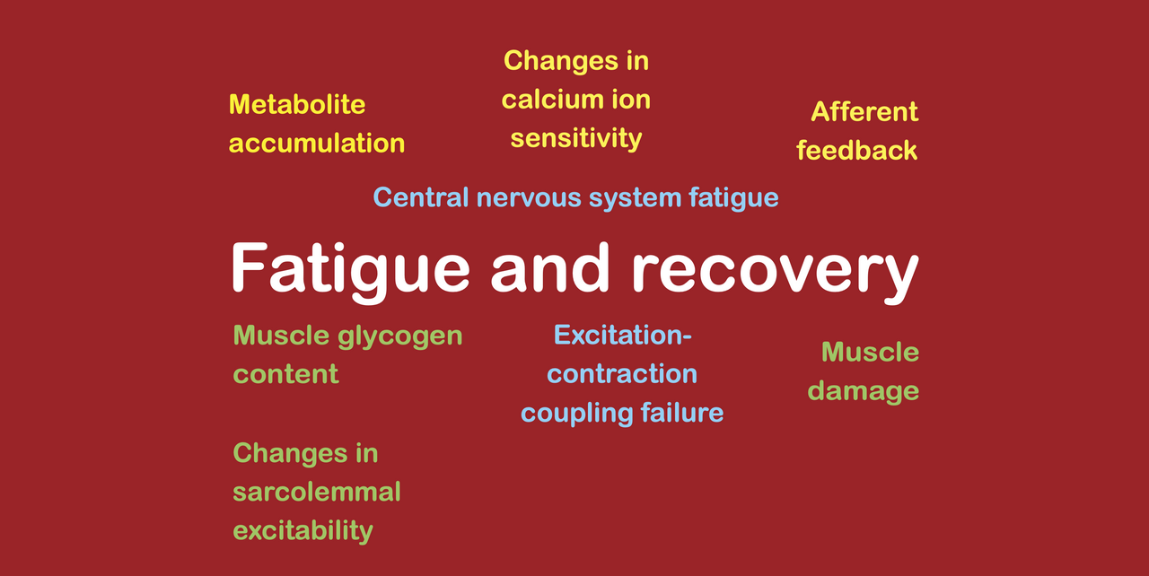
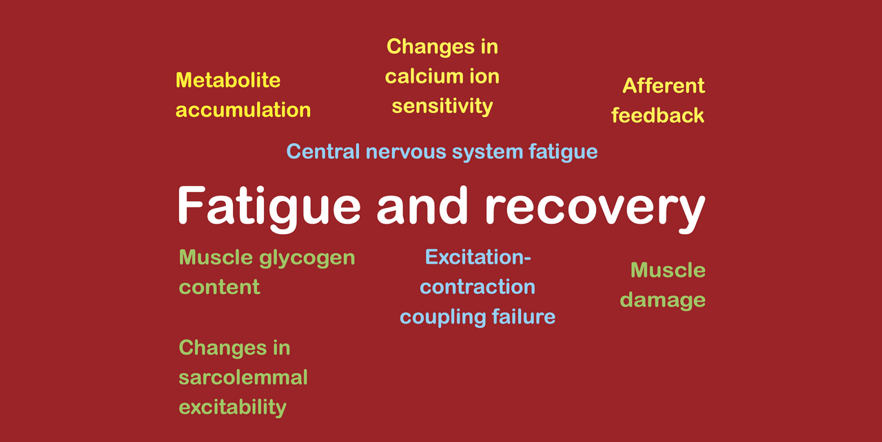
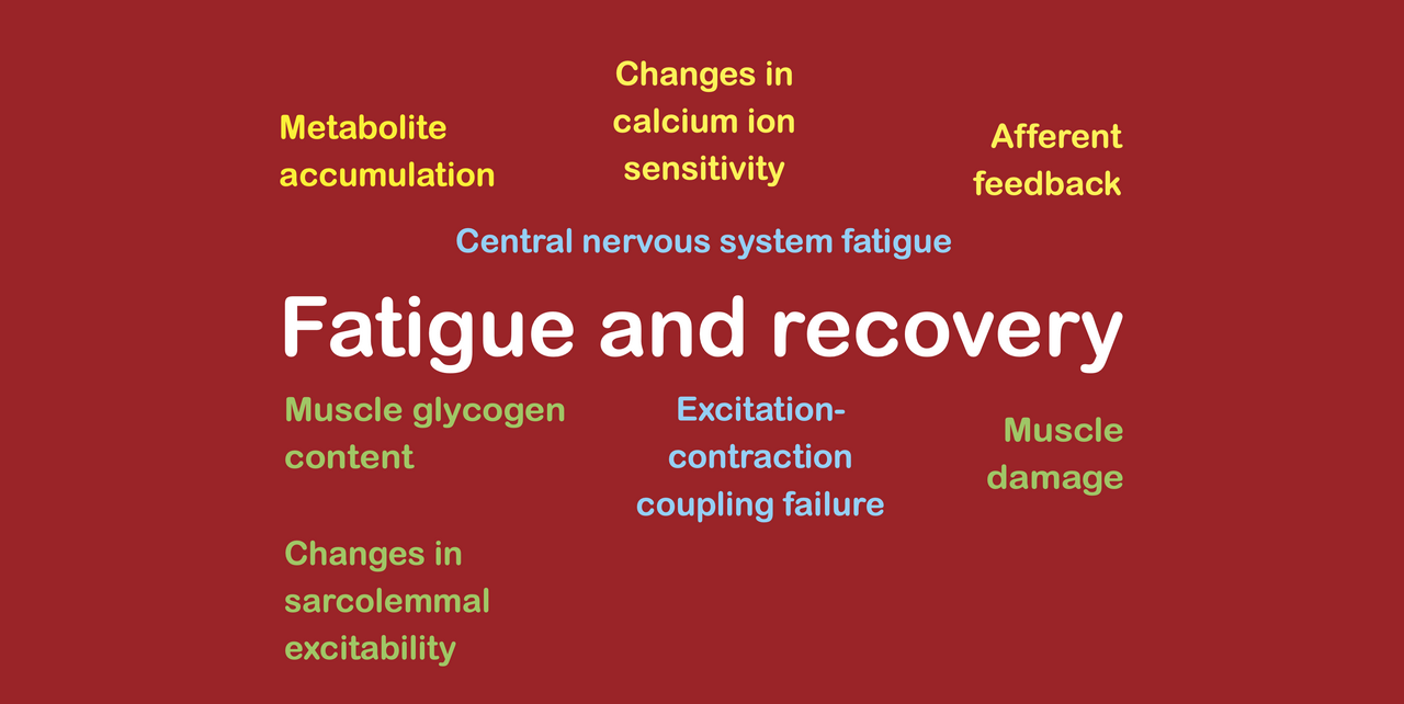
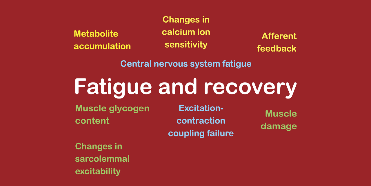
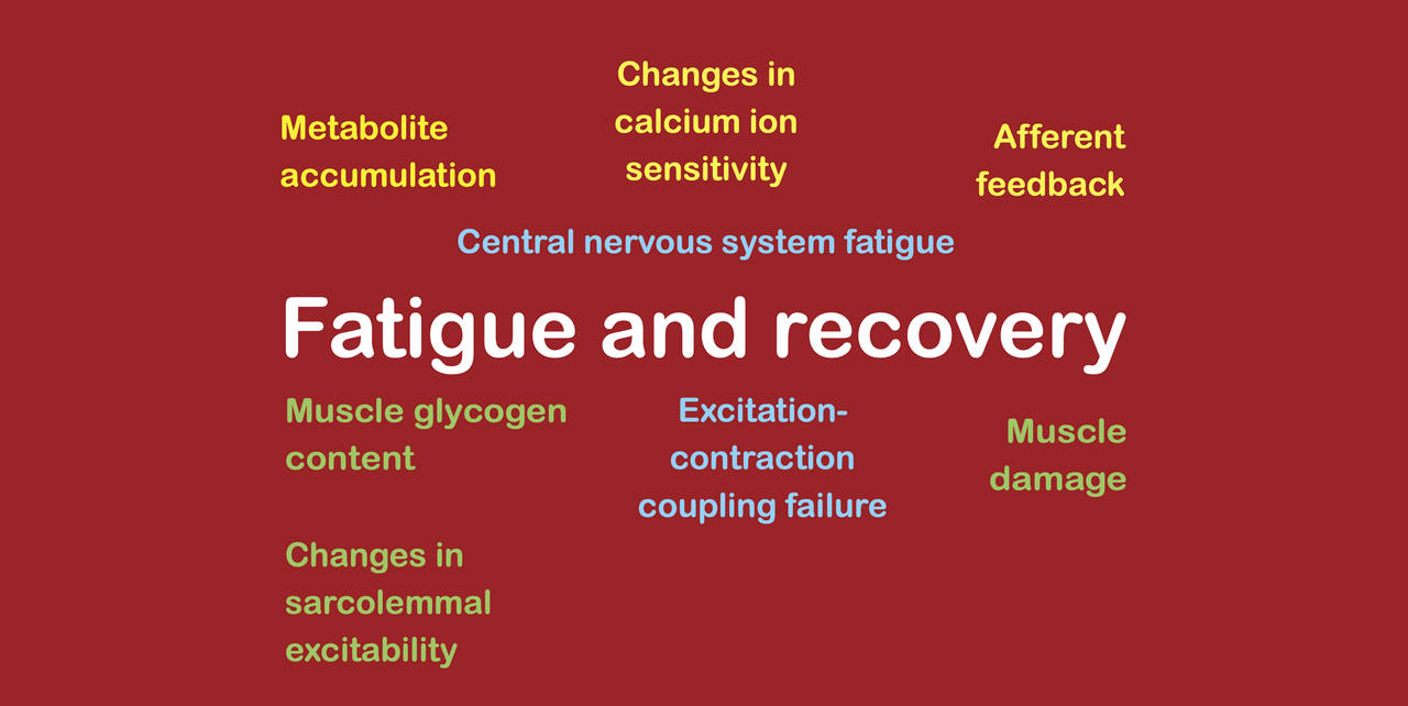
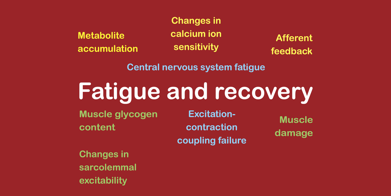
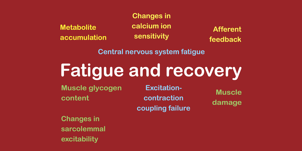
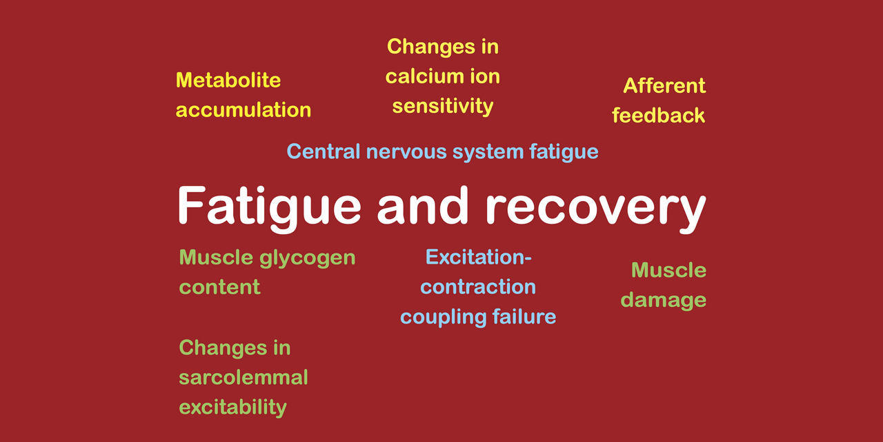
  3. calcium ion accumulation (including excitation-contraction coupling failure, losses of calcium ion sensitivity, and reductions in sarcolemmal excitability);

  4. spinal CNS fatigue; and

  5. supraspinal CNS fatigue (caused by afferent feedback due to metabolite accumulation or inflammatory mediators, and by inflammatory mediators entering the bloodstream).

Importantly, each of these fatigue mechanisms do not all dissipate at the same rate. Some fatigue mechanisms dissipate very quickly, while others dissipate much more slowly. Specifically, metabolite accumulation dissipates within approximately half an hour, glycogen depletion is reversed within a single day (although it likely causes this long-lasting fatigue through calcium ion-related mechanisms rather than directly by the absence of fuel), calcium ion-related fatigue can last for days if it is caused by excitation-contraction coupling failure, spinal CNS fatigue probably dissipates within a couple of minutes, and supraspinal CNS fatigue probably dissipates within half an hour (when it is caused by afferent feedback from metabolite accumulation) or a few hours (if caused by afferent feedback from inflammatory mediators). So the only long-lasting fatigue mechanism that occurs during exercise involves calcium ion accumulation in general and excitation-contraction coupling failure in particular.


What mechanisms of fatigue develop only after a bout of exercise?

#1. Muscle damage

Muscle damage is commonly believed to be generated during the bout of exercise itself. This is because it is assumed to occur as a result of the very high levels of mechanical tension that are present during eccentric contractions stretching muscle fibers until they break (leading to the “popping” of individual sarcomeres) as well as structural damage to the cytoskeleton and the muscle cell membrane. However, many studies have shown that muscle damage is not in fact present immediately after a bout of exercise that involves eccentric contractions. Rather, there is a delay of at least several hours before any signs of sarcomere rearrangement become apparent. How is this possible?

In fact, muscle damage is not actually caused by the high levels of mechanical tension that occur during eccentric contractions stretching muscle fibers until they break. Muscle fibers probably only physically break during repeated stretches if they are either [A] exposed to lengths that are way beyond those that can be achieved physiologically, or [B] excessively fatigued before being actively stretched. And these circumstances are more accurately described as muscle strain injury rather than muscle damage.

Rather, most muscle damage is actually caused by a biochemical effect, resulting from the influx and accumulation of calcium ions into the muscle fiber when muscle activation is sustained during a bout of exercise. When calcium ions are released out of the sarcoplasmic reticulum to stimulate the actin-myosin crossbridge binding process that causes muscle fibers to produce force, they are usually taken back up into the sarcoplasmic reticulum immediately afterwards. However, when contractions are repeated and muscle activation is sustained, calcium ions can accumulate inside the muscle fiber. If the mitochondria do not remove them quickly enough, the presence of these calcium ions then triggers a signaling response leading to the production of calpains (proteases) that degrade the insides of the cell. Researchers have shown that there is a pronounced increase in calpain levels immediately after a bout of muscle-damaging exercise, which is related to the sustained fatigue that is experienced.

While some commentators have assumed that the tendency for eccentric contractions to cause more muscle damage than concentric and isometric contractions is clear proof that mechanical tension is involved in producing muscle damage, this claim totally ignores the fact that eccentric contractions cause the opening of stretch-activated ion channels that allow the influx of additional calcium ions into the muscle fiber, causing a large stimulus for calpain production and therefore both muscle damage and excitation-contraction coupling failure. It is not necessary to resort to the high mechanical tension that is produced during eccentric contractions in order to explain how large amounts of muscle damage are produced.

Indeed, the process that causes muscle damage is clearly similar to the process that causes excitation-contraction coupling failure. In fact, it is identical, except that the calpains attack the minor triadic proteins that lie between the voltage sensor of the transverse tubules and the calcium ion store of the sarcoplasmic reticulum. Therefore, it is valuable to compare and contrast the development of excitation-contraction coupling failure with the development of muscle damage. In this respect, it is noteworthy that while excitation-contraction coupling failure clearly develops fairly rapidly during a bout of exercise (albeit not nearly as rapidly as metabolite accumulation during strength training), muscle damage does not become apparent until hours after a bout of exercise.

Excitation-contraction coupling failure occurs relatively quickly during a bout of exercise, partly because it can probably be temporarily created by other mechanisms and partly because it seems to require very little calpain activity for damage to occur to the minor proteins of this triadic junction such that excitation-contraction coupling failure occurs. In fact, this makes sense, because the triadic junction works by maintaining a close contact between the voltage sensor and the calcium ion store. Even a small disruption to the small, triadic proteins that hold the two structures adjacent to one another can result in the electrical signal that arrives at the voltage sensor failing to trigger the release of calcium ions into the cytoplasm from the calcium ion store. In contrast, muscle damage refers to wholesale restructuring of myofibrils and the cytoskeleton, which are much larger than the minor triadic proteins. Thus, the process of causing damage to them takes longer to play out, and it is not apparent until several hours after the workout has been completed.


#2. Supraspinal central nervous system (CNS) fatigue

While it is true that supraspinal CNS fatigue occurs during exercise, the nature of that supraspinal CNS fatigue is very transitory. Thus, we certainly cannot say that the supraspinal CNS fatigue that is produced during exercise contributes to the supraspinal CNS fatigue that persists several hours and days after a workout has been completed. Nevertheless, supraspinal CNS fatigue can be measured several days after exercise, which means that some other sources of supraspinal CNS fatigue must arise once the workout has finished.

During a strength training set, supraspinal CNS fatigue arises as a result of the afferent feedback from metabolites accumulating inside the muscle. The accumulating metabolites stimulate metaboreceptors that then send information back up afferent nerves to the brain, where it is registered as fatiguing or burning sensations. These perceptions of fatiguing or burning are then added to the perception of effort, which reduces the magnitude of the central motor command that can be attained at the maximal tolerable effort level. This in turn causes supraspinal CNS fatigue, which is an inability to produce the normally maximal level of central motor command and therefore the normally maximal level of motor unit recruitment, despite exerting a maximal effort. A similar phenomenon occurs during aerobic exercise, where supraspinal CNS fatigue arises as a result of the afferent feedback from inflammatory mediators accumulating inside the muscle (noting that these inflammatory mediators also enter the bloodstream and communicate with the brain through this system as well as by means of afferent feedback). Nevertheless, once the metabolites and the inflammatory mediators have dissipated, the supraspinal CNS fatigue similarly goes away. Therefore, this type of supraspinal CNS fatigue can only last for an hour or two after a bout of exercise, at the very most.

Nevertheless, the muscle damage response that occurs once exercise has been completed typically involves an inflammatory response. This inflammatory response is integral to the muscle damage and repair processes. Yet, it also causes supraspinal CNS fatigue in much the same way as the inflammatory mediators cause supraspinal CNS fatigue during aerobic exercise (as well as during long strength training workouts). While it has yet to be shown that these particular inflammatory mediators can cause supraspinal CNS fatigue by means of afferent feedback from the muscles, they have been found to cause supraspinal CNS fatigue by entering the bloodstream and causing increases in perceptions of fatigue and effort by acting directly on the brain. Therefore, the supraspinal CNS fatigue that is observed in the days after a strength training workout or after a bout of aerobic exercise is not caused by the same phenomena as the supraspinal CNS fatigue that was observed during the exercise bouts themselves.


What does this mean in practice?

Given that the only long-lasting fatigue mechanism that occurs during exercise is excitation-contraction coupling failure and given that the only mechanisms of fatigue that occur after exercise are muscle damage and supraspinal CNS fatigue, we can identify these three types of fatigue as being the sole causes of sustained fatigue in the days after a workout. This is important, since muscle damage and excitation-contraction coupling failure are both triggered by the same ultimate cause, which is the accumulation of calcium ions inside a muscle fiber as a result of a sustained period of muscle activation, and supraspinal CNS fatigue is itself caused by the inflammatory response to muscle damage. This means that there is actually only one ultimate cause of the sustained fatigue that is present in the days after a workout, and that is the accumulation of calcium ions during the workout as a result of the sustained period of muscle activation.

Consequently, within the context of a single contraction mode (eccentric, concentric, or isometric), we can now predict the amount of sustained fatigue that a single muscle fiber will experience after a workout based on the duration of time for which it is activated. Moreover, we can also predict the amount of fatigue that a whole muscle will experience after a workout based on the duration of time for which it is activated.

Unfortunately, these relationships between muscle activation time and muscle damage are not linear for either muscle fibers or whole muscles, because calcium ion accumulation seems to remain relatively low (likely while mitochondria are removing the ions) until a certain time point, after which it accelerates fairly quickly. Indeed, this is why proximity to muscular failure affects fatigue during strength training, because it is the final two reps of a set that seem to involve a large increase in excitation-contraction coupling failure. Also, the relationship between the duration of time for which a whole muscle is activated and the resulting fatigue will not be linear because slow twitch muscle fibers are less susceptible to calcium ion accumulation than fast twitch muscle fibers (since they have more mitochondria). Thus, very long workouts or bouts of aerobic exercise during which only slow twitch muscle fibers are active will not cause a huge amount of sustained fatigue, while even relatively short workouts or bouts of strength training during which fast twitch muscle fibers are active have the potential to cause far more sustained fatigue.

Nevertheless, even though the relationships are not linear, we can still say that the primary factor that causes fatigue after a workout is the duration of time for which muscle fibers and whole muscles are activated. This gives us a principle that we can use to minimize the sustained fatigue that occurs, since we can now aim to achieve a stimulus for whatever outcome is required (increased maximum strength, speed, muscle size) by maximizing the stimuli that cause adaptations suitable for those outcomes (increased ability to recruit motor units, increased coordination, increased muscle fiber diameter and length) while simultaneously reducing the duration of time for which muscle fibers are activated. For example, if we consider that there are only five or six stimulating reps per set to muscular failure, then we should be able to minimize fatigue by working with sets of 5–7 reps rather than by working with higher numbers of reps, where the added muscle activation time does not add any incremental benefit but causes much more sustained fatigue to accumulate.


What is the takeaway?

Fatigue is a temporary reduction in exercise performance as a result of previous exercise. During strength training exercise, fatigue is caused by metabolite accumulation, calcium ion accumulation, spinal CNS fatigue, and supraspinal CNS fatigue. During aerobic exercise, fatigue is caused by glycogen depletion, calcium ion accumulation, spinal CNS fatigue, and supraspinal CNS fatigue. After exercise, only excitation-contraction coupling failure (a type of fatigue caused by calcium ion accumulation) persists. However, muscle damage and supraspinal CNS fatigue caused by the inflammatory response subsequent to muscle damage arise in the hours and days following the exercise bout, such that the only three types of fatigue that are present in a sustained way are [1] excitation-contraction coupling failure, [2] muscle damage, and [3] supraspinal CNS fatigue. Moreover, since excitation-contraction coupling failure and muscle damage are caused by calcium ion accumulation subsequent to muscle activation, and since supraspinal CNS fatigue is caused by the inflammatory response to muscle damage, all three sustained fatigue mechanisms are ultimately caused by the exposure of muscle fibers to a sustained period of activation. Therefore, minimizing muscle activation duration while maximizing the key stimuli that cause adaptations is the key to optimizing strength training frequency.
Please subscribe to my newsletter to read the future articles in this series!


Fatigue is a temporary reduction in exercise performance as a result of previous exercise. Since it is temporary, the mechanisms that cause fatigue during exercise dissipate, leading to a recovery of strength. However, at the same time as some fatigue mechanisms dissipate, others develop in a delayed fashion (in the same way as delayed onset muscle soreness).

To understand fatigue in the hours and days following a workout, we therefore need to understand [A] how the fatigue mechanisms that arise during exercise dissipate, and [B] how the fatigue mechanisms that develop after exercise appear and then dissipate again. In this article, I am going to focus solely on the dissipation of those fatigue mechanisms that actually develop during exercise, and in a subsequent article I will focus on the delayed fatigue mechanisms that only appear after exercise.


What mechanisms of fatigue develop during a bout of exercise?

Fatigue is a temporary and reversible reduction in exercise performance due to a preceding bout of exercise. During exercise, fatigue can arise through several different fatigue mechanisms, including mechanisms inside the central nervous system (CNS), which lead to reductions in the level of central motor command that reaches the muscle (thereby reducing motor unit recruitment and motor unit firing rates), and mechanisms inside the muscle itself, which lead to reductions in either muscle fiber force or muscle fiber shortening velocity. Considering all kinds of exercise (including strength training and other forms of anaerobic exercise as well as endurance exercise) we can identify several groups of fatigue mechanisms, as follows:

  1. metabolite accumulation;

  2. glycogen depletion;

  3. calcium ion accumulation;

  4. spinal CNS fatigue; and

  5. supraspinal CNS fatigue.

Most of these groups themselves include several individual fatigue mechanisms. For example, metabolite accumulation involves both acidosis and phosphate ion accumulation; glycogen depletion can occur in multiple different locations within a muscle fiber (within myofibrils, between myofibrils, and close to the sarcolemma); calcium ion accumulation causes a range of fatigue mechanisms, including excitation-contraction coupling failure, reduced calcium ion sensitivity, and reduced sarcolemmal excitability; and supraspinal CNS fatigue can be caused by a range of different types of afferent feedback, including from metabolites in the working muscles and inflammatory mediators in the bloodstream. Each of these individual fatigue mechanisms is itself caused by slightly different factors, has a slightly different effect, and develops and dissipates at a different speed. For this reason, it is helpful to consider each mechanism carefully if we want to understand the way in which fatigue dissipates after a bout of exercise.


How does each mechanism of fatigue dissipate after a bout of exercise?

Metabolite accumulation

Metabolite accumulation causes fatigue through at least two separate mechanisms, being acidosis (the accumulation of hydrogen ions) and the accumulation of phosphate ions. Acidosis primarily reduces muscle fiber shortening velocity by acting on the rate at which myosin heads attach and detach from actin myofibrils, while the presence of phosphate ions reduces muscle fiber force by preventing the supply of ATP from being turned into ADP and a phosphate ion plus energy for the crossbridge cycle. Importantly, the types of fatigue that are caused by both acidosis and the presence of phosphate ions are only apparent when hydrogen ions and phosphate ions are present. As soon as these ions are removed, the fatigue is similarly immediately removed. Both hydrogen ions and phosphate ions can freely exit out of the muscle fiber when blood flow is not occluded by intramuscular or extramuscular pressure. Thus, as soon as a bout of exercise is finished, blood is able to flow freely into the muscle and remove the metabolites, which typically occurs very quickly. Most of the accumulated metabolites are able to leave a muscle within a few minutes and the muscle is back to its resting levels within half an hour. For this reason, metabolite accumulation is one of the most rapidly dissipating groups of fatigue mechanisms.

Glycogen depletion

Glycogen depletion is often assumed to cause fatigue directly by means of reducing the supply of available fuel. However, it actually seems to cause peripheral fatigue predominantly by impairing the release of calcium ions from the sarcoplasmic reticulum (likely by means of intramyofibrillar glycogen depletion contributing to excitation-contraction coupling failure) and it may also cause supraspinal CNS fatigue by afferent signaling of progressively reducing fuel supplies. Thus, the way in which glycogen depletion causes fatigue is probably not by means of its own fatigue mechanism. Instead, it works by stimulating other fatigue mechanisms. When glycogen is depleted during non-damaging exercise, it typically takes 24 hours or so to recover. During the period of time when glycogen is depleted, fatigue can develop more rapidly during exercise, and this is most likely because the presence of low glycogen levels exacerbates the development of excitation-contraction coupling failure and supraspinal CNS fatigue.

Calcium ion accumulation

INTRODUCTION

Calcium ion accumulation inside muscle fibers occurs as a result of repeated muscle activation. As a muscle fiber is activated, this stimulates the release of calcium ions from its sarcoplasmic reticulum into the cytoplasm, where the ions stimulate the binding of myosin heads to actin myofibrils, to form the crossbridges that generate muscle fiber force. As the muscle fiber deactivates (activation and deactivation occur many times per second), the calcium ions are pulled back into the sarcoplasmic reticulum before the process repeats itself again. However, the process is not perfect, and every time calcium ions are released and then pulled back into the sarcoplasmic reticulum, some of them are left behind in the cytoplasm. This means that free calcium ions gradually accumulate inside the muscle fiber, and this causes at least three different calcium ion-related fatigue mechanisms: excitation-contraction coupling failure, a reduction in sarcolemmal excitability, and a loss of myofibrillar sensitivity.

EXCITATION–CONTRACTION COUPLING FAILURE

Excitation-contraction coupling failure occurs when the calcium ions build up around the triadic junction between the voltage sensor and the sarcoplasmic reticulum calcium ion store. When they accumulate in this area, they stimulate the release of proteases that degrade the minor proteins that hold the triadic junction in place. After the junction destabilizes, the voltage sensor pulls away from the sarcoplasmic reticulum calcium ion store, which stops the arrival of the electrical signal along the cell membrane from causing the release of calcium ions from the sarcoplasmic reticulum. Importantly, since this process involves damage, neither the removal of the calcium ions nor even the removal of the proteases can cause immediate recovery. Recovery only occurs after the muscle fiber has been able to repair the damage to the triadic junction, which can take several days.

REDUCTION IN SARCOLEMMAL EXCITABILITY

A reduction in sarcolemmal excitability occurs when the calcium ions build up inside the cytoplasm near to the mitochondria, and are removed from the fiber by them. When this happens, the mitochondria release reactive oxygen species into the cytoplasm. These reactive oxygen species stimulate the release of phospholipases that subsequently cause small holes in the muscle cell membrane. When the cell membrane is damaged in this way, it reduces the extent to which the electrical signal can travel along the cell membrane. Importantly, since this process involves damage, neither the removal of the calcium ions, nor the removal of the reactive oxygen species, nor the removal of the phospholipases can cause immediate recovery. Recovery only occurs after the muscle fiber has been able to repair the holes in the muscle cell membrane, which seems to take several hours under normal circumstances.

LOSS OF MYOFIBRILLAR SENSITIVITY

A loss of myofibrillar sensitivity occurs when the actin myofibrils inside the muscle fiber stop responding to the presence of calcium ions. Exactly why this happens is still unclear. However, when it does happen, the myosin heads stop binding to actin myofibrils even when there are calcium ions present inside the cytoplasm. It is possible that the removal of the excessive amounts of calcium ions from the cytoplasm can cause a rapid reversal of this fatigue mechanism, such that it does not last for a long period of time after exercise, but there is still comparatively little research in this area in comparison with the other calcium ion-related fatigue mechanisms.

Spinal CNS fatigue

Spinal CNS fatigue during exercise appears to be caused by the rapid and repetitive firing of motor neurons. Indeed, recent research has shown that only those motor neurons that fire during a bout of exercise are fatigued at the spinal level (in this way spinal CNS fatigue differs from supraspinal CNS fatigue, which affects all motor neurons and does so starting from the high-threshold motor units downwards, even when those motor units are not actually used during exercise). Although there is relatively little research that is able to differentiate between spinal and supraspinal CNS fatigue during and after single bouts of exercise, it does seem quite likely that spinal CNS fatigue vanishes within a couple of minutes after stopping exercise. This is because there is a very sudden reduction in overall CNS fatigue within the first couple of minutes of stopping exercise, and it cannot easily be attributed to the loss of supraspinal CNS fatigue caused by afferent feedback, since the underlying causes of this afferent feedback do not dissipate that rapidly.

Supraspinal CNS fatigue

Supraspinal CNS fatigue during exercise is caused mainly by afferent feedback from the working muscles and cardiovascular system. Supraspinal CNS fatigue refers to the inability of the motor cortex to generate a sufficiently large neural signal (called the “central motor command”) to send to the working muscle and produce maximum motor unit recruitment. During exercise, supraspinal CNS fatigue seems to be caused predominantly by afferent feedback that raises our perceived level of effort without a concomitant increase in the magnitude of the central motor command.

How does this work? Well, to produce a movement, the motor cortex generates a central motor command. The larger the size of the central motor command, the more motor units are recruited, and the more force can be produced. While the central motor command goes primarily to the muscle to recruit motor units, it also sends a copy of itself (called the “corollary discharge”) to another part of the brain, where we register it as a perception of effort. When the central motor command is large, we perceive a high level of effort, and when the central motor command is small, we only perceive a low level of effort. For this reason, when we reach a maximum tolerable level of effort, we cannot increase our central motor command any further, because doing so would simultaneously increase our perceived level of effort. In this way, our maximum tolerable level of effort is the limiting factor on motor unit recruitment levels at any moment in time. Consequently, if any other factors increase our perception of effort (such as afferent feedback sources), then this increases our perceived level of effort faster than our central motor command, causing us to reach our maximum tolerable level of effort at a lower level of central motor command. When this happens, we necessarily experience supraspinal CNS fatigue.

Afferent feedback during strength training comes primarily from the accumulation of metabolites. When metabolites accumulate in large amounts throughout the whole muscle (and not just inside muscle fibers), they activate metaboreceptors, which send information back up afferent nerves to the brain. We perceive this information in the sensory parts of our brains as the presence of burning and fatiguing sensations located inside the muscle. Importantly, this information increases our perception of effort, thereby causing supraspinal CNS fatigue. Similar effects are caused by the inflammatory mediators that are released by the muscle and cause more generalized fatiguing sensations during long durations of exercise (whether strength training or aerobic exercise).

Obviously, when supraspinal CNS fatigue is caused by the presence of afferent feedback, it will dissipate once the substances that are causing the afferent feedback also dissipate. Consequently, supraspinal CNS fatigue during strength training that is caused by metabolite accumulation will necessarily disappear after a few minutes. Similarly, supraspinal CNS fatigue during long durations of exercise that is caused by the appearance of inflammatory mediators in the muscles and the blood will probably also disappear within a couple of hours (albeit the exact timing its disappearance is still unclear).


What does this mean in practice?

In practice, with the exception of calcium ion-related fatigue mechanisms (including the effects that are downstream of glycogen depletion), all fatigue mechanisms that arise during exercise probably dissipate within a couple of hours after exercise. This means that the fatigue that is present on the day after a workout must necessarily be caused by either the calcium ion-related fatigue that was incurred in the workout itself or by additional fatigue mechanisms that develop after the workout was completed. Interestingly, as we will find out in the next article, all of these additional fatigue mechanisms are actually stimulated by calcium ion-related processes as well. As a result, this means that any fatigue mechanism that is present hours or days after a bout of exercise must be the result of calcium ion-related mechanisms.


What is the takeaway?

Fatigue is a temporary reduction in exercise performance as a result of previous exercise. Since they are temporary, the mechanisms that cause fatigue during exercise necessarily dissipate after the workout has been completed, leading to a recovery of strength. Nevertheless, these fatigue mechanisms do not all dissipate at the same rate. Some fatigue mechanisms dissipate very quickly, while others dissipate much more slowly. Specifically, metabolite accumulation dissipates within approximately half an hour, glycogen depletion is reversed within 24 hours (although it likely causes this long-lasting fatigue through calcium ion-related mechanisms rather than directly by the absence of fuel), calcium ion-related fatigue can last for several days if it is caused by excitation-contraction coupling failure, spinal CNS fatigue probably dissipates within a couple of minutes, and supraspinal CNS fatigue probably dissipates within half an hour (when it is caused by afferent feedback from metabolite accumulation) or a few hours (if caused by afferent feedback from inflammatory mediators). Thus, the only really long-lasting fatigue mechanisms that occur during exercise are those that relate to calcium ion accumulation.

Please subscribe to my newsletter to read the future articles in this series!


Fatigue is a temporary reduction in exercise performance as a result of previous exercise. We can analyze the development of fatigue over the course of a single bout of exercise. Recently, I explained how fatigue develops over a strength training set. In this article, explain exactly how fatigue develops over the course of a bout of aerobic exercise.


What mechanisms of fatigue develop during a bout of aerobic exercise?

Introduction

Fatigue is a temporary and reversible reduction in exercise performance due that is usually caused by preceding exercise. During exercise, fatigue can arise through several different fatigue mechanisms, including mechanisms inside the central nervous system (CNS), which lead to reductions in the level of central motor command that reaches the muscle (thereby reducing motor unit recruitment and motor unit firing rates), and mechanisms inside the muscle itself, which lead to reductions in either muscle fiber force or muscle fiber shortening velocity. To understand how fatigue develops over the course of a bout of aerobic exercise, we need to understand exactly how each of these mechanisms takes effect.


The unique features of aerobic exercise

Aerobic exercise differs from strength training in two key respects. Firstly, it involves lower whole muscle forces, and secondly it involves a much longer duration in which muscles are active. These two key features cause fatigue to develop in very different ways. The fact that whole muscle forces are lower is important because metabolite accumulation cannot occur inside a muscle unless whole muscle forces exceed a certain threshold (which is usually 25–30% of maximum isometric force). Therefore, metabolite-related fatigue does not occur during aerobic exercise. For this reason, the only peripheral fatigue mechanisms are those that involve calcium ion accumulation. And the fact that muscles are activated during aerobic exercise for longer than during a strength training set means that the depletion of glycogen stored inside muscle fibers can have a fatiguing effect.

Ultimately, therefore, we can categorize the various fatigue mechanisms that are working during a bout of aerobic exercise into four groups, as follows: [1] glycogen depletion, [2] calcium ion accumulation, [3] spinal CNS fatigue, and [4] supraspinal CNS fatigue. Let’s now take a look at each of these fatigue mechanisms in more detail.


#1. Glycogen depletion

INTRODUCTION

In general, fatigue mechanisms work by reducing the ability of muscle fibers to complete the necessary steps that lead to force production. For example, supraspinal CNS fatigue stops the motor cortex from creating a sufficiently large central motor command such that the high-threshold motor units can be recruited. Similarly, spinal CNS fatigue reduces the size of the central motor command that is transmitted to the neuromuscular junction, stopping certain motor units from being recruited or reducing the motor unit firing rates of the activated muscle fibers. Calcium ion-related fatigue mechanisms stop the electrical signal that is propagated along activated muscle fibers from turning into a chemical signal that triggers crossbridge formation. And finally, metabolite-related fatigue either slows or stops the crossbridge cycle from occurring by interfering directly with the binding of myosin heads to the actin myofilaments. All of these fatigue mechanisms are therefore impairments in the normal process that leads to muscle fibers producing force. Contrary to popular belief, in none of these fatigue mechanisms does a lack of fuel lead to a reduction in force production or power output. Thus, the idea that fatigue typically involves running out of energy is greatly mistaken. Nevertheless, when we consider endurance exercise, a reduction in the availability of glycogen can cause fatigue to occur.

GLYCOGEN DEPLETION

During sustained, intense aerobic exercise, muscle and liver glycogen levels are reduced as the glycogen is broken down to provide fuel for the muscle fiber. The muscle fiber needs fuel to power the crossbridge cycle as well as to power the excitation-contraction coupling process. Thus, glycogen stores are located in several key places throughout muscle fibers, including inside the myofibrils (intramyofibrillar), between myofibrils (intermyofibrillar glycogen), and next to the sarcolemma (subsarcolemmal glycogen). Intensity and duration of exercise can affect the extent and also the exact location of the glycogen depletion (both in terms of which muscle fiber types are affected and also in terms of where in the muscle fiber the depletion occurs). When intensity is high, both fast twitch muscle fibers and slow twitch muscle fibers are activated, and since fast twitch muscle fibers are less oxidative and more glycolytic, they use proportionally more glycogen, and thereby deplete glycogen at a faster rate. Conversely, when duration is long, there is a longer period of time in which glycogen can be depleted, and since glycogen is not replenished (in either the muscles or the liver) at the same rate as it is used, the longer an exercise bout persists, the greater the potential for glycogen depletion to occur.

FATIGUING EFFECTS OF GLYCOGEN DEPLETION

It is widely assumed that glycogen depletion causes fatigue simply by stopping the delivery of glucose to the muscle fiber such that it can no longer power the crossbridge cycle or excitation-contraction coupling failure. Leaving aside the fact that the complete depletion of muscle fiber glycogen could be quite damaging for a muscle fiber, the majority of the evidence supporting this idea is actually circumstantial, insofar as most of the landmark studies merely show a strong association between exercise performance and levels of muscle glycogen. They do not demonstrate that the mechanism underpinning this association is a failure to provide fuel or a reduction in the supply of fuel. Also, there are at least two other mechanisms by which glycogen depletion could cause fatigue, neither of which involve a reduction in the supply of energy.

Firstly, several investigations have identified links between the availability of muscle glycogen and [A] the ability of the sarcoplasmic reticulum to release calcium ions in response to an electrical signal arriving at the voltage sensor of the triadic junction despite the presence of an alternative fuel source (ATP and phosphocreatine), and [B] fatigue, again despite the presence of an alternative fuel source (ATP and phosphocreatine). Therefore, it seems likely that reduced muscle fiber glycogen in fact causes calcium ion-related fatigue mechanisms rather than acting directly on muscle fiber function by limiting the amount of energy available for crossbridge formation or excitation-contraction coupling.

Secondly, it is feasible that the depletion of muscle glycogen levels during aerobic exercise produces afferent feedback to the brain in much the same way as afferent feedback is produced by the accumulation of metabolites during strength training sets. Indeed, baseline glycogen levels have been shown to affect the pacing strategies employed during endurance activities, suggesting that there is a feedback loop involving the brain and the level of glycogen available inside the muscle. This suggests that reduced muscle fiber glycogen in fact causes supraspinal CNS fatigue rather than acting directly on muscle fiber function by limiting the energy available for either crossbridge formation or excitation-contraction coupling.


#2. Calcium ion accumulation

INTRODUCTION

During a bout of aerobic exercise, calcium ions gradually accumulate inside the working muscle fibers as they are repeatedly activated, and this causes the development of several calcium ion-related peripheral fatigue mechanisms. All of the calcium ion-related fatigue mechanisms are caused by the same basic process, which arises due to repeated muscle activation. Muscle activation is caused by the recruitment of motor units, which in turn is produced by the central motor command sent to the muscle by the motor cortex.

When the motor cortex sends a central motor command to the muscle, it recruits motor units. Each of these motor units controls multiple muscle fibers, and the recruitment of the motor unit causes the immediate activation of every single muscle fiber connected to that motor unit. When a muscle fiber is activated, an electrical signal propagates along its outer membrane and goes down inside the transverse tubules that are situated along its length. At the bottom of a transverse tubule, the electrical signal reaches a voltage sensor located at a junction with the sarcoplasmic reticulum. The junction permits an interaction between the voltage sensor and the sarcoplasmic reticulum, such that the arrival of an electrical signal at the voltage sensor causes the sarcoplasmic reticulum to deposit calcium ions into the cytoplasm. When actin myofibrils detect the presence of calcium ions inside the cytoplasm, they change their conformation, which allows myosin heads to bind with them, and this causes actin-myosin crossbridges to form, which is what generates a muscle fiber force.

As soon as a muscle fiber is activated, it immediately deactivates itself and waits for the next electrical signal from the CNS. When the muscle fiber is deactivated, the electrical signal disappears from the voltage sensor, and this causes the calcium ions to be pulled back into the sarcoplasmic reticulum. Since a muscle fiber is switched on and off many times per second during any muscular contraction, the process of putting calcium ions into the cytoplasm of a muscle fiber and removing them again also occurs many times per second. Nevertheless, not all of the calcium ions that are placed into a muscle fiber are successfully pulled back into the sarcoplasmic reticulum every single time, and this leads to calcium ion accumulation inside the cytoplasm. The accumulation of calcium ions stimulates the release of calpains (which are proteases) and phospholipases that cause various peripheral fatigue mechanisms.

Mitochondria inside the muscle fiber act as the first line of defense against the calcium ions inside the cytoplasm. When they detect calcium ions, they act to remove them. Yet, they can only remove a finite amount of calcium ions and once they are full, the calcium ions are allowed to accumulate freely. The behavior of mitochondria explains why calcium ion-related fatigue mechanisms occur more slowly inside slow twitch muscle fibers than inside fast twitch muscle fibers. Slow twitch muscle fibers have more mitochondria, and hence prevent calcium ion accumulation from occurring as quickly. This is also why calcium ion-related fatigue develops at a slower rate during aerobic exercise than during a strength training set. Only the slow twitch muscle fibers of a muscle are actually working during a bout of aerobic exercise while both slow and fast twitch muscle fibers work during a strength training set.

There are three primary fatigue mechanisms that are stimulated by the presence of accumulating calcium ions inside the cytoplasm, which are: [1] excitation-contraction coupling failure (ECCF), [2] reduced calcium ion sensitivity, and [3] reduced sarcolemmal excitability. ECCF and reduced sarcolemmal excitability appear to share a common mechanistic pathway that involves the stimulation of proteases or lipases by the accumulation of calcium ions inside the cytoplasm, while reduced calcium ion sensitivity probably works slightly differently. Even so, they all basically produce the same effect, which is to stop the electrical signal that arrives at the neuromuscular junction from triggering the formation of crossbridges inside the muscle fibers. In this way, calcium ion-related fatigue mechanisms directly impair muscle fiber force of the activated, working muscle fibers.

EXCITATION–CONTRACTION COUPLING FAILURE (ECCF)

The accumulation of calcium ions inside the cytoplasm stimulates the release of proteases called calpains. These calpains degrade the minor triadic proteins that hold the triadic junction in place. When this happens, the voltage sensor drifts away from the sarcoplasmic reticulum calcium ion store, such that the two structures can no longer interact with each other. Subsequently, when an electrical signal reaches the bottom of a transverse tubule and interacts with the voltage sensor, this fails to stimulate a release of calcium ions into the cytoplasm from the sarcoplasmic reticulum calcium ion store. Since the conversion of the electrical signal into the chemical signal at the triadic junction is called “excitation-contraction coupling,” we refer to this fatigue mechanism as a failure of excitation-contraction coupling or ECCF.

REDUCED SARCOLEMMAL EXCITABILITY

The accumulation of calcium ions inside the cytoplasm additionally stimulates the release of phospholipases, although the process by which these particular lipases are generated is probably slightly more complex than the process in which calpains are produced. Indeed, phospholipases are likely produced in response to the production of reactive oxygen species (ROS) that are created by mitochondria when they handle excess calcium ions. Thus, the generation of phospholipases as a result of the presence of calcium ions is slightly more indirect. Nevertheless, it is still the accumulation of calcium ions that causes the phospholipases to be produced. These phospholipases degrade the muscle fiber cell membrane. When this happens, the electrical signal that normally propagates along the surface of the cell membrane is impaired, such that it fails to reach the transverse tubules. Subsequently, when a muscle fibers is activated, the electrical signal does not propagate properly. Since the muscle cell membrane is also called the “sarcolemma” and since the membrane allows the transmission of electrical signals due to its polarity, we refer to this fatigue mechanism as a loss of sarcolemmal excitability.

REDUCED CALCIUM ION SENSITIVITY

The accumulation of calcium ions inside the cytoplasm also causes a reduction in the sensitivity of actin myofibrils to the presence of calcium ions. In other words, the actin myofibrils stop responding to the presence of calcium ions in the cytoplasm. This calcium ion-related peripheral fatigue mechanism is the least well-known of the three main calcium ion-related fatigue mechanisms. While it seems likely that it occurs due to the presence of a signaling molecule inside the cytoplasm, the exact mechanism is still unclear.


#3. Spinal central nervous system (CNS) fatigue

In order to produce muscular contractions, the motor cortex generates a central motor command that is transmitted down the spinal cord to the neuromuscular junction of the muscle, where it recruits motor units (and those motor units subsequently activate groups of muscle fibers). This central motor command is sent repeatedly by the brain down the spinal cord to the muscle, many times per second. The repeated transmission of the signal down the spinal cord gradually produces an impairment of the magnitude of the signal that reaches the other end. Over time, it becomes apparent that a smaller signal is emerging at the neuromuscular junction despite a similar (or often even larger) signal being created by the motor cortex. This impairment of the magnitude of the central motor command as it is transmitted down the spinal cord is defined as spinal CNS fatigue.

Although the mechanisms that cause spinal CNS fatigue are still very unclear, it possesses two very important features. Firstly, the duration of time during which the signal is transmitted is what determines the magnitude of the spinal CNS fatigue. Therefore, short durations of exercise cause little spinal CNS fatigue, and the magnitude of the spinal CNS fatigue that is experienced by the working muscle fibers increases with exercise duration. Secondly, it is only the working motor units (and their linked muscle fibers) that experience spinal CNS fatigue. As we will see in the subsequent section, this means that spinal CNS fatigue is very different in nature from supraspinal CNS fatigue.


#4. Supraspinal central nervous system (CNS) fatigue

INTRODUCTION

In order to produce muscular contractions, the motor cortex generates a central motor command that is transmitted down the spinal cord to the neuromuscular junction of the muscle, where it recruits motor units (and those motor units subsequently activate groups of muscle fibers). The size of the central motor command determines the number of motor units that are recruited and therefore the number of muscle fibers that are activated. When we are unfatigued, the motor cortex can produce a very large central motor command, such that we achieve a high level of motor unit recruitment, and most of the muscle fibers in a muscle are activated. However, if we experience supraspinal CNS fatigue, the level of central motor command that the motor cortex can produce is reduced, such that fewer motor units are recruited and fewer muscle fibers are activated.

Importantly, since supraspinal CNS fatigue occurs inside the motor cortex, it affects the size of the central motor command that is generated and not the quality of the central motor command that arrives at the neuromuscular junction. The size of the central motor command determines how many motor units are recruited, with larger size signals allowing the recruitment of more (and higher threshold) motor units. For this reason, although supraspinal CNS fatigue might arise due to the activation of only the slow twitch muscle fibers of low-threshold motor units during aerobic exercise, its effect is primarily to reduce the access to the fast twitch muscle fibers of high-threshold motor units (starting with the highest high-threshold motor units and gradually progressing down the line until eventually the slow twitch muscle fibers are reached, which is what ultimately disrupts exercise performance).

Supraspinal CNS fatigue is measured by assessing voluntary activation by the interpolated twitch technique, which compares the force produced during voluntary and involuntary contractions in unfatigued and fatigued states, to isolate the loss in voluntary (not involuntary) force experienced as a result of the fatiguing exercise bout. To understand how this supraspinal CNS fatigue arises, we have to understand how the central motor command that the motor cortex can produce is influenced by [A] our perceived level of effort, and [B] by our current level of motivation.

#1. PERCEIVED LEVEL OF EFFORT

When we produce a muscular contraction by creating a central motor command signal, this signal automatically also generates a secondary signal called the corollary discharge. This secondary signal travels to another part of the brain where it is detected and recognized as a key contributor to our perception of effort. Thus, whenever we create a central motor command signal, we automatically also experience a perception of effort. Moreover, the larger the central motor command signal, the larger the perceived level of effort. Since our ability to tolerate perceived effort has a maximum threshold, the corollary discharge acts as a negative feedback loop to produce a cap on the size of the central motor command signal that we can achieve. Once we reach our maximum tolerable perceived effort level, we are prevented from increasing the size of our central motor command signal. In practice, this means that anything else (other than the corollary discharge) that increases our perceived level of effort will reduce our ability to reach a truly maximum level of central motor command.

The presence of afferent feedback from the body is one such factor that can increase our perceived level of effort. During aerobic exercise, inflammatory mediators are slowly released into muscles and also into the bloodstream, and this transmits afferent feedback to the brain both directly through receptors inside the muscles as well as by means of the circulatory system. As these inflammatory mediators increase our level of perceived effort, they cause us to approach our maximum tolerable level of perceived effort without increasing the level of central motor command. Thus, as the amount of inflammatory mediators increases, the maximum possible level of central motor command that we can achieve decreases, which means that supraspinal CNS fatigue occurs and is measurable by way of reductions in voluntary activation. Since the supraspinal CNS fatigue is dependent upon the gradual and incremental release of inflammatory mediators, it increases over time. Therefore, longer durations of aerobic exercise cause more supraspinal CNS fatigue.

#2. MOTIVATION

Our maximum tolerable level of perceived effort varies slightly from one day to the next and from one moment to the next during a bout of exercise as a result of our motivation levels. This level of motivation can alter depending on how we perceive the task that we are performing. If we feel positive and confident about our achievement, we will likely be able to tolerate a higher level of perceived effort. In contrast, if we feel negative and disillusioned about our current performance, we will typically be less able to tolerate discomfort. Thus, our motivation acts as a multiplier for our maximum tolerable level of perceived effort, and therefore either expands or contracts the capacity of the motor cortex to produce a central motor command. The malleable nature of motivation and its subsequent impact on our maximum tolerable level of perceived effort can explain some otherwise very puzzling phenomena that occur during endurance exercise tasks, such as the second wind and the ability to achieve a finishing sprint. No central governor is actually needed, only an understanding of how our motivation is affected by our processing of the situation and the subsequent effects of that motivation level on our maximum tolerable level of perceived effort.

We say that supraspinal CNS fatigue is present during a bout of exercise if the levels of central motor command that the motor cortex can produce during that bout of exercise are suppressed in comparison with baseline levels. Yet, the way in which supraspinal CNS fatigue is measured during bouts of aerobic exercise do not really take the effects of the exercise bout on motivation into account. Indeed, supraspinal CNS fatigue is usually measured in this context by stopping the aerobic exercise briefly and performing a short, maximal effort strength test, while simultaneously recording voluntary activation using the interpolated twitch technique. The problem with this approach is that the level of motivation required to exert a very short, maximal effort (while taking a break from the long distance endurance event) is much smaller than the level of motivation required to continue exerting a sustained effort over a very long period of time. For this reason, supraspinal CNS fatigue induced by changes in motivation is probably greatly underestimated during aerobic exercise.


Practical implications

WHAT CAUSES TASK FAILURE DURING AEROBIC EXERCISE?

Aerobic exercise performances are typically measured by way of either time trials or races. In such situations, the athletes attempt to achieve the highest possible level of sustained power output for the required time or distance, in order to achieve the longest distance in the time provided, or finish the set distance in the shortest possible time. As explained above, various fatigue mechanisms act to reduce this power output, and all of them increase their contributions over time.

Inside the working muscle fibers, three calcium ion-related mechanisms (ECCF, reductions in sarcolemmal excitability, and reductions in calcium ion sensitivity) reduce the muscle fiber force that working muscle fibers can exert, by stopping the muscle fiber from responding to the presence of the electrical signals being sent to them. Glycogen depletion likely also reduces the extent to which muscle fibers produce force, although it may actually exert its effects peripherally predominantly by exacerbating the calcium ion-related fatigue (the impact of reduced fuel availability is likely overstated). Simultaneously, spinal CNS fatigue occurs that impairs the size of the signal being sent to these working muscle fibers. This reduces the level of muscle fiber force exerted (by reducing motor unit firing frequency) and may also stop the muscle fibers from being activated at all, if the size of the electrical signal drops below a key threshold. These mechanisms are all duration-dependent, such that the fatiguing effects increase progressively over time.

In response to these peripheral fatigue mechanisms, the athlete must try to increase the size of the central motor command being sent to the working muscles, to recruit additional motor units (and hence also additional muscle fibers) that compensate for the fatigue inside the currently working muscle fibers. In this way, fatigue instigates the same voluntary response during aerobic exercise as it does during strength training sets.

Nevertheless, at the same time as the peripheral fatigue mechanisms are occurring, the athlete experiences supraspinal CNS fatigue due to reducing motivation (which reduces the maximum tolerable level of perceived effort) as well as due to increasing afferent feedback (likely due to the accumulation of inflammatory mediators and the depletion of muscle fiber glycogen). These mechanisms reduce the maximum possible level of central motor command that can be achieved at any given moment in time. Thus, the ability to achieve a given level of exercise performance is squeezed between an increasing level of peripheral (and spinal CNS) fatigue that demands an ever increasing magnitude of central motor command to be generated by the motor cortex, and an increasing level of supraspinal CNS fatigue that reduces the level of central motor command that can be attained. When these two mechanisms meet in the middle, the ability to maintain the existing power output suddenly disappears and the athlete is said to hit the wall (although in reality, if changes in motivation are achievable in this situation, the wall might be temporarily lifted, since this elevates the maximum tolerable level of perceived effort and allows power output to be maintained for a slightly longer time).


What is the takeaway?

During a bout of aerobic exercise, various fatigue mechanisms develop over time, including [1] glycogen depletion, [2] calcium ion accumulation, [3] spinal CNS fatigue, and [4] supraspinal CNS fatigue. As the peripheral fatigue types develop, the athlete must voluntarily increase effort levels to recruit additional motor units and thereby activate more muscle fibers in compensation. Yet, the developing supraspinal CNS fatigue simultaneously reduces the number of motor units that can be recruited, ultimately leading to task failure being reached when the two types of fatigue mechanism meet in the middle.

Please subscribe to my newsletter to read the future articles in this series!


If you have read anything about the science of hypertrophy in recent years, you will have come across the idea that motor unit recruitment levels increase in tandem with increasing fatigue, during strength training sets that involve a self-selected tempo or a slow, fixed tempo (as is very common in bodybuilding). The levels of motor unit recruitment that are experienced during a rep are important, because it is the level of motor unit recruitment that determines how many muscle fibers are activated, and therefore trained. Thus, the higher the level of motor unit recruitment during a rep, the greater the number of muscle fibers that are trained, and the more hypertrophy the rep is able to stimulate (although the exact magnitude of the stimulus depends on the level of mechanical tension, which varies independently of whether a muscle fiber is activated or not). Therefore, in this article, I plan to explain exactly how fatigue interacts with levels of motor unit recruitment in each rep over the course of a strength training set.


What is motor unit recruitment?

When we want to perform a muscular contraction in order to create a movement, we first generate a signal inside the motor cortex of the brain, which is called the central motor command. The size of this central motor command is set according to how fast we want the weight to move. Larger central motor commands are created when we want to move a weight as fast as we possibly can, while smaller central motor commands are created when we want to move a weight comparatively more slowly.

The central motor command is then sent to two places. Firstly, it is sent to another part of the brain, where it registers as a perception of effort. This means that we have immediate feedback of our intention to produce a large effort, in the form of a perception. Secondly, it is sent to the intended muscle, where it recruits motor units. Each motor unit is a group of muscle fibers (although these muscle fibers are not grouped closely together, but are in fact spread out throughout a specific region of the muscle). When a single motor unit is recruited, all of its muscle fibers are immediately activated. The size of the central motor command determines how many motor units are recruited. Larger central motor commands recruit larger numbers of motor units, and smaller central motor commands recruit smaller numbers of motor units.

Importantly, while activating more muscle fibers allows more muscle fibers to contribute to whole muscle force, simply activating a single muscle fiber does not guarantee that it also produces a high force and therefore experiences the high level of mechanical tension that is necessary to stimulate hypertrophy. The force produced (and experienced) by each individual muscle fiber is determined mainly by the force-velocity relationship, the length-tension relationship, and the extent of peripheral fatigue. We can consider muscle fiber activation as essentially “switching a muscle fiber on” while these other factors determine the mechanical tension that the activated muscle fiber then experiences. Indeed, this is why very fast movements (like jumping and throwing) do not stimulate hypertrophy despite being performed with very high levels of muscle activation.


What determines motor unit recruitment levels?

Although it is common to see strength training commentators refer to motor unit recruitment levels increasing as a result of fatigue, this is not strictly accurate. Central motor commands are generated entirely voluntarily, and therefore they cannot be either created or increased by the presence of fatigue (or by any other factor, for that matter). Additionally (and most importantly), the magnitude of the central motor command depends on how fast we intend to move the weight.

If we are unfatigued and we want to lift a light weight with a very slow tempo, our intention to move in this way will necessarily create a small central motor command (because this small central motor command in turn generates a fairly low level of motor unit recruitment, and therefore only activates a small number of muscle fibers, which is sufficient for the low level of force that is needed to create the small acceleration necessary for a slow bar speed). Conversely, if we are unfatigued and we want to move the same weight as fast as possible, we will necessarily create a large central motor command (because this generates a high level of motor unit recruitment and therefore activates a large number of muscle fibers, which are necessary for the high level of force that is needed to create a large acceleration that is required in order to achieve the very fast movement speed).


What determines effort levels?

Effort during a strength training set is typically measured by the level of perceived effort or the rating of perceived exertion (RPE). Traditionally, the RPE was defined as a self-reported scale for the subjective sensation of work that is naturally associated with any muscular contraction.

As explained above, the central motor command sends a signal to a sensory part of the brain that creates a subjective sensation of work associated with the muscular contraction that is being performed (this signal is called the corollary discharge). In this way, the central motor command directly contributes to the perception of effort. Thus, when central motor command is low, the perception of effort will also often (but not always) be low. In contrast, when central motor command is high, the perception of effort will always be high. The reason that the perception of effort is not always low when the central motor command is low is because other factors can additionally increase the perception of effort as well as the central motor command.

We can now include the perception of effort into the previous example.

If we are unfatigued and we want to lift a light weight with a very slow tempo, our intention to move in this way will necessarily create a small central motor command (because this small central motor command in turn generates a fairly low level of motor unit recruitment, and therefore only activates a small number of muscle fibers, which is sufficient for the low level of force that is needed to create the small acceleration necessary for a slow bar speed). In addition, the small central motor command will also produce a low perceived level of effort or RPE. Conversely, if we are unfatigued and we want to move the same weight as fast as possible, we will necessarily create a large central motor command (because this generates a high level of motor unit recruitment and therefore activates a large number of muscle fibers, which are necessary for the high level of force that is needed to create a large acceleration that is required in order to achieve the very fast movement speed). In addition, the large central motor command will also produce a high perceived level of effort or RPE. This is why we can refer to throwing a ball as fast as possible as being the same thing as throwing a ball with a maximal effort.

Hopefully, by this point it should be clear that fatigue has absolutely nothing to do with either motor unit recruitment levels or effort levels during a muscular contraction. Both are entirely under voluntary control, and are necessarily both under voluntary control, otherwise we would be unable to gauge how much force to produce in order to create appropriate movements.


What is the limit for the maximum level of motor unit recruitment in a muscular contraction?

Typically, it is not possible for us to access all of the motor units in a muscle during a muscular contraction. Moreover, the number of motor units in a muscle that can be accessed varies between individuals (more well-trained lifters are able to access more motor units than less-well trained individuals), between muscles (smaller muscles permit access to access more motor units than larger muscles), and state of fatigue (generally, more motor units can be accessed when fatigue is totally absent compared to when it is present).

Since the number of motor units that are recruited during a muscular contraction is dependent upon the magnitude of the central motor command, if we were able to continue increasing the size of the central motor command without any external limitation, then we would be able to achieve complete motor unit recruitment. As this is not possible, some other factor must be limiting the magnitude of the central motor command that we can create during a muscular contraction.

It seems likely that the limit is the perceived level of effort.

The perception of effort is discomforting. Therefore, we tend to try and avoid experiencing high levels of effort, especially for sustained periods of time. Therefore, we probably have a maximal tolerable level of perceived effort that we are unwilling or unable to exceed (albeit this threshold does probably vary according to our transient motivation levels for the task in hand). The perceived level of effort is increased by the corollary discharge signal created by the central motor command (as well as other factors). Thus, any increase in the central motor command also creates an increase in the level of perceived effort. Since the level of perceived effort has a maximum tolerable level, this also imposes an upper limit on the maximum possible level of central motor command (although again, this depends on the context).


What other factors increase effort levels (and why does this matter)?

While the corollary discharge signal sent by the central motor command to another part of the brain is the main contributing factor for our perceived level of effort, other factors can also contribute to the perception of effort. Such factors increase the perceived level of effort in the same way as the corollary discharge (although without similarly increasing the central motor command). This is important because they cause our perception of effort to reach its maximum tolerable level at a lower level of central motor command. This then causes the maximum possible level of central motor command to be lower, compared to when these other factors are not present.

There are two main factors that increase the perceived level of effort in a strength training set in this way: [1] metabolite accumulation, and [2] the presence of inflammatory mediators. They both seem to work in essentially the same way.

As metabolites accumulate inside a muscle during a strength training set, they stimulate metaboreceptors inside the muscle. These metaboreceptors send afferent feedback to the brain, where they create an increase in the perceived level of effort, alongside burning and fatiguing sensations. While these sensations are probably what contribute to the increased level of perceived effort, they do not also increase the central motor command. Therefore, they cause us to reach our maximum tolerable level of perceived effort at a lower level of central motor command than if they were not present.

Similarly, as inflammatory mediators accumulate inside a muscle during a strength training set, they stimulate other receptors inside the muscle. These receptors send afferent feedback to the brain, where they create an increase in the perceived level of effort, alongside generalized sensations of fatigue. In addition, the inflammatory mediators also enter the bloodstream and can be detected across the blood-brain barrier (from where it also creates generalized sensations of fatigue). In this way, inflammatory mediators can exert effects on perceived effort through two pathways, both of which contribute to an increased level of perceived effort without also increasing the central motor command. Therefore, like accumulating metabolites, they similarly cause us to reach our maximum tolerable level of perceived effort at a lower level of central motor command than if they were not present.

In practice, this means that the presence of metabolites and the presence of inflammatory mediators both reduce the maximum central motor command that can be sent to the muscle. This is essentially supraspinal central nervous system (CNS) fatigue. We are involuntarily prevented from creating the level of central motor command that we would normally be able to attain, and thus we are similarly involuntarily prevented from creating the level of motor unit recruitment that we would normally be able to attain. Clearly, this means that when metabolite accumulation is high (as during light load strength training to failure or when using certain advanced techniques), maximum motor unit recruitment levels will be slightly reduced. Similarly, when inflammatory mediators are present (such as towards the end of a long workout), maximum motor unit recruitment levels will also be reduced, and probably to a greater extent than when metabolites are present.

Importantly, this reduction in the maximum level of motor unit recruitment during exercise then prevents us from achieving gains in the ability to recruit high-threshold motor units in the future (since it is the very high level of central motor command that acts as the stimulus for this adaptation). And it may also slightly reduce the amount of hypertrophy that can be stimulated, since fewer muscle fibers are being trained.


How do recruitment and effort change over the course of a strength training set?

As I explained in my earlier article, peripheral fatigue mechanisms occur over the course of a strength training set. When some of the muscle fibers within a muscle experience peripheral fatigue, they necessarily stop being capable of producing the required force or shortening at the required velocity. For this reason, unless additional muscle fibers are activated to compensate for this reduction in performance (by means of an increase in central motor command leading to an increase in motor unit recruitment level), the bar speed being used during the set will slow down.

Strength training sets can be performed in one of two main ways. Either they can be performed with maximal intended bar speed on every rep (as is the case during velocity-based training) or they can be performed with a self-selected or a slow fixed tempo on every rep.

When a maximal intended bar speed is used on every rep, central motor command (and therefore motor unit recruitment) and the level of perceived effort are both maximal on the first couple of reps of the set. Thereafter, central motor command gradually decreases slightly to its lowest level at the end of the set, whether the set is terminated before reaching muscular failure (as is often the case with velocity-based training) or whether the set is carried out until muscular failure. However, the perception of effort will remain at a maximal level on all reps of the set. The gradual reduction in central motor command occurs mainly due to the accumulation of metabolites, which slowly starts to increase the afferent feedback, leading to an increase in perceived effort due to mechanisms unrelated to the central motor command. For this reason, central motor command must reduce to compensate and maintain maximum tolerable levels of effort at the same level. In addition, the central motor command that is transmitted to the muscle reduces over time, as a result of spinal CNS fatigue (wherein repeated motor neuron firing reduces the extent to which the signal is transmissible down the spinal cord). Thus, contrary to popular belief, the presence of fatigue actually impairs the level of motor unit recruitment during this type of strength training.

When a self-selected tempo or a fixed slow tempo are used on every rep, central motor command (and therefore motor unit recruitment) and the level of perceived effort are both relatively low on the first couple of reps of the set. Thus, not all muscle fibers are activated. Nevertheless, as peripheral fatigue accumulates inside the working muscle fibers, they experience reductions in their ability to support the movement at the intended velocity. For this reason, additional muscle fibers must be activated to compensate, or else tempo will slow down even further. To maintain the intended tempo, the lifter voluntarily chooses to increase central motor command, which in turn increases the number of recruited motor units, which in turn increases the number of activated muscle fibers. This process increases over the course of the set until the maximum central motor command reaches its maximal level towards the end of the set, whether the set is terminated before reaching muscular failure or whether the set is carried out until muscular failure. Nevertheless, given that metabolite accumulation and inflammatory mediators also build up in this set, and given that spinal CNS fatigue also occurs, the maximum level of central motor command (and the maximum level of motor unit recruitment) in the set is not as high as when maximal efforts are used from the first rep of the set. This is why using maximal intended bar speed on every rep of a set is important for maximizing strength gains.


What is the takeaway?

Fatigue has nothing to do with either motor unit recruitment levels or effort levels during a muscular contraction. Both are entirely under voluntary control. Indeed, it is necessary that both are under voluntary control, otherwise we would be unable to gauge how much force to produce in order to create appropriate movements. Nevertheless, the presence of fatigue during a strength training set can influence the level of motor unit recruitment that is achieved. During strength training sets that involve a maximal intended bar speed on every rep, the presence of peripheral fatigue impairs motor unit recruitment levels, by sending afferent feedback to the brain and thereby creating supraspinal CNS fatigue (such that maximal motor unit recruitment is achieved on the first rep and minimal motor unit recruitment is attained on the final rep). In contrast, during strength training sets with a self-selected or slow fixed tempo, the presence of peripheral fatigue prompts the lifter to use a greater effort, which in turn increases the level of motor unit recruitment to its maximal level on the final rep (which is still lower than the levels achieved during strength training with a maximal intended bar speed on every rep).

Please subscribe to my newsletter to read the future articles in this series!


Fatigue is a temporary reduction in exercise performance as a result of previous exercise. We can analyze the development of fatigue over the course of a single bout of exercise. In my previous article, I explained exactly how fatigue develops over the course of a normal strength training set. In this short follow-up article, I clarify how weight on the bar (and therefore the number of repetitions taken to reach muscular failure) affects which types of fatigue are most prevalent.


How does fatigue develop over a set, and why are the individual mechanisms important?

Introduction

Strength training often involves lifting a weight repeatedly until muscular failure. Muscular failure occurs whenever sufficient fatigue is present such that the muscle can no longer produce enough force, even at a very slow velocity, in order to lift the weight. This fatigue can arise due to many different fatigue mechanisms, including mechanisms inside the central nervous system (CNS), which lead to reductions in the level of central motor command that reaches the muscle (thereby reducing motor unit recruitment and motor unit firing rates), and mechanisms inside the muscle itself.


How fatigue develops over a strength training set

During a normal strength training set, various fatigue mechanisms develop over time. Together, these mechanisms reduce the ability of the muscle to produce sufficient force at the required bar speed, and ultimately lead to muscular failure being reached.

The first fatigue mechanism to appear is acidosis, which predominantly reduces muscle fiber shortening speed. This allows single muscle fiber force to remain high despite an increase in effort (and motor unit recruitment) being needed in order to maintain self-selected tempo. Later, calcium ion-related mechanisms also appear that reduce single muscle fiber force, which in turn reduces the magnitude of single muscle fiber mechanical tension. In addition to metabolite-related fatigue and calcium ion-related fatigue, spinal and supraspinal central nervous system (CNS) fatigue increase progressively over the course of a strength training set. These reduce both the level of motor unit recruitment that can be attained (which obviously reduces whole muscle force being produced, essentially by switching off groups of muscle fibers) and the motor unit firing rates that are attained (which reduces single muscle fiber force of the working muscle fibers).

Ultimately, the gradual accumulation of all of these fatigue mechanisms causes muscular failure to be reached. However, what is important for our purposes today is that the relative contributions of each of these types of fatigue differs depending on the weight that is being lifted.


How does load affect how fatigue develops over a set?

Introduction

Although the mainstream fitness industry tends to assume that metabolite-related fatigue is more important during light load strength training to failure and CNS fatigue is more important during heavy load strength training, this is actually the wrong way around. In fact, as we will see, metabolite-related fatigue contributes more to overall fatigue when lifting heavy loads (even though the total amount of metabolites that accumulate is indeed smaller), and CNS fatigue contributes more to overall fatigue when lifting light loads. To appreciate why this might be the case, let’s look more closely at the fatigue mechanisms involved in lifting heavy loads and light loads. Indeed, this analysis is very much worth doing, because it helps us explain why heavy load strength training and light load strength training produce somewhat different adaptations, at least in terms of gains in maximum strength.


Fatigue when lifting heavy loads

When lifting heavy loads (which are greater than or equal to 5RM), muscular failure is reached very quickly. Heavy loads always require maximal efforts and therefore full motor unit recruitment, which means that all of the fast twitch (highly glycolytic) muscle fibers are activated from the first rep of the set. These muscle fibers quickly accumulate metabolites inside them and therefore progress very rapidly through the acidosis and inorganic phosphate phases of metabolite-related fatigue, such that they quickly get to the point where they are unable to contribute to whole muscle force. Since the force produced by these muscle fibers is necessary for the weight to be lifted, muscular failure is essentially reached by the localized fatigue of a small number of muscle fibers (and the heavier the weight, the fewer muscle fibers are in fact fatigued).

In this situation, metabolite-related fatigue is absolutely critical to the attainment of muscular failure when lifting heavy loads, even though the overall muscle does not accumulate a large amount of metabolites. It is only the accumulation of a small amount of metabolites in the fast twitch muscle fibers of the highest high-threshold motor units that causes muscular failure. Indeed, this is one of those situations in which we realize that the terminology “muscular failure” is somewhat misleading because the failure to achieve the task of lifting a heavy load is actually brought about solely by the fatigue of a very small number of muscle fibers.

The lack of a meaningful accumulation of metabolites throughout the whole muscle when lifting heavy loads has an important corollary effect, which is that very little afferent feedback is generated. Thus, contrary to popular belief, the amount of supraspinal CNS fatigue is actually very low during heavy load strength training exercise.

Additionally, it is important to note that the very rapid attainment of muscular failure when lifting heavy loads reduces the contribution of those fatigue mechanisms that depend upon the duration of time for which muscle fibers are activated. Thus, all of the calcium ion-related mechanisms have very little opportunity to contribute, since they require time to accumulate and to stimulate the actions of calpains. Similarly, the contribution of spinal CNS fatigue is much reduced, because it depends upon the repeated firing of the motor neurons in order to take effect.

Overall, we can therefore appreciate that metabolite-related fatigue is the most important type of fatigue during heavy load strength training exercise, even though whole muscle metabolite accumulation is still fairly low. The levels of supraspinal CNS fatigue are low (because whole muscle metabolite accumulation is low). Similarly, levels of spinal CNS fatigue and calcium ion-related fatigue mechanisms are also low, because of the short duration of time for which the working muscles are active. Indeed, this is an important reason why heavy loads are very effective for achieving the largest gains in maximum strength, because the low levels of spinal and supraspinal CNS fatigue permit very high levels of motor unit recruitment to be achieved at muscular failure, which in turn stimulates large gains in the ability to recruit high-threshold motor units in future workouts or strength tests.


Fatigue when lifting light loads

When lifting light loads, muscular failure takes a long time to be achieved. To reach the point at which the whole muscle is unable to exert the force to lift the weight, a large proportion of the fibers inside the muscle must be greatly fatigued. This has two important implications.

Firstly, it means that the total amount of metabolites that accumulate inside the muscle is very substantial (which leads to a large amount of afferent feedback, which we detect as the fatiguing and burning sensations inside the muscle, and which causes a large amount of supraspinal CNS fatigue).

Secondly, it means that the time for which all of the working muscle fibers are active is much longer, which allows sufficient time for calcium ions to accumulate (and therefore to stimulate the actions of calpains and cause calcium ion-related fatigue mechanisms and it also increases the extent of spinal CNS fatigue, which is also time dependent, because it relies upon the repeated firing of the motor neurons in order to take effect.

Thus, in contrast to lifting heavy loads, light load strength training to failure involves much more CNS fatigue (both spinal and supraspinal) and more calcium ion-related fatigue. The high levels of CNS fatigue during sets explain why training with light loads is much less effective for achieving gains in maximum strength, because the high levels of spinal and supraspinal CNS fatigue at muscular failure reduce the maximum achievable levels of motor unit recruitment, which in turn then fails to stimulate gains in the ability to recruit high-threshold motor units.


What is the takeaway?

During a normal strength training set, various fatigue mechanisms develop over time, which together ultimately lead to a reduction in the ability of the lifter to perform the exercise, leading to muscular failure. Importantly, the load used greatly affects the types of fatigue that are experienced. Heavier loads involve a proportionally much greater contribution from metabolites (even though metabolite accumulation is less) and this means that they involve low levels of calcium ion-related fatigue and low levels of CNS fatigue. Since they involve low levels of CNS fatigue, they permit extremely high levels of central motor command at muscular failure, which is how they are able to cause the greatest gains in the ability to recruit high-threshold motor units. Conversely, training with lighter loads involves much more calcium ion-related fatigue and CNS fatigue, which is why they involve smaller gains in the ability to recruit high-threshold motor units.

Please subscribe to my newsletter to read the future articles in this series!


Fatigue is a temporary reduction in exercise performance as a result of previous exercise. We can analyze the development of fatigue over the course of a single bout of exercise. In this article, I hope to explain exactly how fatigue develops over the course of a normal strength training set.


What mechanisms of fatigue develop during a normal strength training set?

Introduction

Strength training often involves lifting a weight repeatedly until muscular failure. There are many misconceptions about exactly what muscular failure involves, and I will clarify exactly how the process of reaching muscular failure works in a future article. Indeed, exactly what muscular failure means is very important for understanding how strength training works, because proximity to failure has been revealed as a key training variable that affects muscular adaptations, particularly hypertrophy. Nevertheless, for the purposes of this article, we can proceed on the basis that muscular failure occurs whenever sufficient fatigue is present such that the muscle can no longer produce enough force, even at a very slow velocity, in order to lift the weight. This fatigue can arise due to many different fatigue mechanisms, including mechanisms inside the central nervous system (CNS), which lead to reductions in the level of central motor command that reaches the muscle (thereby reducing motor unit recruitment and motor unit firing rates), and mechanisms inside the muscle itself.


The unique feature of strength training

Since conventional strength training involves lifting at at least a light load (if not a moderate or even a heavy load), each muscular contraction must involve a force that is higher than 25% of maximum whole muscle force. This is a very important point, because this level of whole muscle force is sufficient to cause venous occlusion for as long as the muscle is activated. While venous occlusion is present, the blood cannot leave the muscle while it is contracting, unlike with lower levels of whole muscle force. For this reason, conventional strength training differs from many other types of exercise because it permits metabolite accumulation to occur, and this facilitates the appearance of metabolite-related fatigue mechanisms. Such metabolite-related fatigue mechanisms are not present when lower whole muscle forces are used (such as is often the case during aerobic exercise), because the lack of venous occlusion allows the blood to flow out of the muscle even while it is contracting, and the blood takes the accumulating metabolites with it.


Similarities between strength training and other exercise

In addition to metabolite-related fatigue, other fatigue mechanisms are also possible during a strength training set, since the repeated activation of the muscle fibers causes calcium ion accumulation inside the cytoplasm, which leads to calcium ion-related fatigue mechanisms, and the repeated firing of the motor neurons leads to spinal central nervous system (CNS) fatigue. Additionally, as the environment inside the muscle changes and receptors detect these alterations, they send afferent signals to the brain that generate supraspinal CNS fatigue. Unlike metabolite-related fatigue, these fatigue mechanisms are also shared with all other types of exercise, including endurance exercise.


The difference between CNS fatigue and peripheral fatigue

It is important to note that the peripheral fatigue mechanisms relate only to those single muscle fibers that are working (in contrast, those muscle fibers that are not activated by a muscular contraction do not experience peripheral fatigue), and affect either their maximum muscle fiber shortening velocity or their maximum muscle fiber force. In contrast, the CNS fatigue mechanisms work by reducing central motor command, which reduces motor unit recruitment levels (thereby switching off the muscle fibers belonging to the highest threshold motor units, irrespective of whether they were working at the time) and which also reduces motor unit firing frequency (thereby reducing the muscle fiber force of all muscle fibers, again irrespective of whether they were working at the time). Thus, while we talk about fatigue mechanisms in general terms, the peripheral fatigue mechanisms always only affect the working muscle fibers, while the CNS fatigue mechanisms mainly affect the muscle fibers of high-threshold motor units. While this point is not particularly important for the current analysis, it becomes essential to take into account whenever we want to clarify the effects of different strength training tempos, techniques, and methods on the way in which fatigue develops.


Fatigue mechanisms during a strength training set

Since metabolite-accumulation occurs during strength training, and both calcium ion-related fatigue mechanisms and CNS fatigue mechanisms occur during all types of exercise, we can categorize the various fatigue mechanisms that are working during a strength training set into four groups, as follows: [1] metabolite accumulation, [2] calcium ion accumulation, [3] spinal CNS fatigue, and [4] supraspinal CNS fatigue. Let’s now take a look at each of these fatigue mechanisms in more detail.


#1. Metabolite accumulation

INTRODUCTION

During a normal strength training set, metabolites accumulate. The two primary metabolites are hydrogen ions (acidosis) and inorganic phosphates. When the muscular contractions of a normal strength training set involve venous occlusion (which happens either when whole muscle force exceeds 25% of maximum or when blood flow restriction is implemented), these metabolites cannot leave the muscle during the set, and often even remain in the muscle fiber in which they originated. Therefore, they accumulate, and this is what causes their fatigue mechanisms to occur.

ACIDOSIS

The accumulation of hydrogen ions produces a fatigue mechanism with two very important characteristics. Firstly, it exerts fatiguing effects very quickly on the muscle fibers that it affects, and therefore it is probably the first mechanism to influence exercise performance during a strength training set. Hence, when we are trying to understand the effects of fatigue mechanisms in the early part of a strength training set, we can focus primarily on acidosis. Secondly, acidosis primarily acts on the attachment and detachment rates of the actin-myosin crossbridges inside the muscle fibers, with the result that it reduces their maximum muscle fiber shortening velocities. Therefore, when we are considering the effects of acidosis, we need to take into account that it primarily reduces exercise performance in the strength training set by reducing muscle fiber shortening speed and not by reducing muscle fiber force (and therefore mechanical tension).

PHOSPHATES

Conversely, the accumulation of inorganic phosphate takes longer to produce an effect, and it exerts its effects on the supply of energy into the crossbridge cycle. For myosin heads to bind repeatedly with actin and form crossbridges to generate force, they require a steady supply of ATP. The reaction that provides energy to the crossbridge cycle involves breaking ATP down into ADP and an inorganic phosphate. As this reaction occurs multiple times, the amount of ADP and phosphate ions accumulating inside the cytoplasm increases. And since the reaction is reversible, the accumulation of phosphate ions eventually suppresses the ability of ATP to break down and produce additional phosphate ions. This causes crossbridge formation to grind to a halt, which in turn eventually reduces muscle fiber force (and hence also mechanical tension). Importantly, since it is the presence of too many phosphate ions that stops additional phosphate ions being produced, the breakdown of ATP can be stopped even in the presence of an existing supply of ATP (and intramuscular glycogen). Thus, we need to be aware that this fatigue mechanism does not involve running out of fuel, but rather the refusal of the actin-myosin motors to accept any more fuel due to the presence of unwanted byproducts.


#2. Calcium ion accumulation

INTRODUCTION

During a normal strength training set, calcium ions accumulate and cause peripheral fatigue mechanisms. In some ways, the accumulation of calcium ions might be perceived as similar to the accumulation of metabolites, but there are important differences in terms of the way that the fatigue mechanisms are produced. Firstly (unlike metabolites), calcium ions do not accumulate inside the cytoplasm due to the metabolic processes that provide energy to the crossbridge cycle. Instead, they are placed into (and then removed from) the cytoplasm by the sarcoplasmic reticulum in response to the repeated activation of the muscle fiber. Secondly (unlike metabolites), calcium ions cannot easily leave the cytoplasm, so it does not matter whether venous occlusion is achieved during each of the muscular contractions in the set (which means that calcium ion-related fatigue occurs even when whole muscle force is less than 25% of maximum force).

Calcium ions accumulate inside muscle fibers because the process that places them into the cytoplasm and then removes them again is not entirely perfect. Electrical signals propagate along muscle cell membranes and go down inside the transverse tubules, where they interact with the sarcoplasmic reticulum at the triadic junction. This interaction causes the sarcoplasmic reticulum to deposit calcium ions into the cytoplasm, which then trigger crossbridges to form. Whenever the electrical signal stops, the calcium ions are pulled back into the sarcoplasmic reticulum. Since the muscle fiber is switched on and off many times per second during any muscular contraction, the process of putting calcium ions into the cytoplasm of a muscle fiber and removing them again also occurs many times per second. Nevertheless, not all of the calcium ions that are placed into a muscle fiber are successfully pulled back into the sarcoplasmic reticulum, and this leads to calcium ion accumulation. Some of these accumulating calcium ions are removed by mitochondria (and this delays the onset of calcium ion-related fatigue, especially in the more oxidative, slow twitch muscle fibers that have more mitochondria), but eventually the ions accumulate, and this is what leads to calcium ion-related fatigue mechanisms.

There are three primary calcium ion-related fatigue mechanisms, which are: [1] excitation-contraction coupling failure (ECCF), [2] reduced calcium ion sensitivity, and [3] reduced sarcolemmal excitability. ECCF and reduced sarcolemmal excitability appear to share a common mechanistic pathway that involves the stimulation of proteases or lipases by the accumulation of calcium ions inside the cytoplasm, while reduced calcium ion sensitivity probably works slightly differently.

EXCITATION–CONTRACTION COUPLING FAILURE (ECCF)

At a high level, ECCF occurs when the electrical signal that propagates along the cell membrane and reaches the transverse tubules fails to cause the release of calcium ions into the cytoplasm from the sarcoplasmic reticulum. Normally, excitation-contraction coupling occurs when the electrical signal being propagated along the cell membrane goes down into the transverse tubules and reaches the voltage sensor at the triadic junction, where it interacts with the sarcoplasmic reticulum calcium ion store to trigger the release of those calcium ions into the cytoplasm. These calcium ions in turn signal to actin and myosin myofibrils to form crossbridges that generate force.

At a more granular level, ECCF occurs when the two sides of the triadic junction drift apart from one another, such that the arrival of the electrical signal at the voltage sensor fails to interact with the sarcoplasmic reticulum calcium ion store. This appears to occur when excess calcium ions inside the cytoplasm stimulate the release of calpains, which degrade the minor proteins that hold the triadic junction in place. The calcium ions accumulate inside the cytoplasm after repeated entry and removal due to the excitation-contraction coupling process itself. Indeed, when a batch of calcium ions is dropped into the cytoplasm from the sarcoplasmic reticulum, all of them are not always taken back up into the sarcoplasmic reticulum. The ones remaining are left to accumulate, and if they are not removed by mitochondria, they stimulate the release of calpains.

Therefore, the process of ECCF involves several steps and so it takes time to appear, in contrast to metabolite-related fatigue mechanisms, which are much faster. In practice, ECCF appears to occur just in time for the last two reps of a strength training set with moderate loads (albeit it probably appears slightly earlier when training with lighter loads and slightly later when training with heavier loads, which is a key point that we will return to when considering the effects of load on fatigue both during and after a workout).

REDUCED SARCOLEMMAL EXCITABILITY

Reduced sarcolemmal excitability involves a very similar mechanism to ECCF, except it is the release of reactive oxygen species (ROS) that stimulates the release of phospholipases that damage the inside of the cell membrane, which in turn prevents the propagation of the electrical signal along the muscle fiber surface. These ROS are stimulated to appear by several mechanisms, including the actions of mitochondria in collecting any calcium ions that accumulate inside the cytoplasm subsequent to excitation-contraction coupling. When this mechanism stops the propagation of the electrical signal along the muscle fiber surface, the level of muscle activation is reduced despite the presence of a strong central motor command reaching the neuromuscular junction. Like ECCF, the process of reducing sarcolemmal excitability involves several steps and takes time to appear, in contrast to metabolite-related fatigue. In practice, reduced sarcolemmal excitability may in fact only become apparent several minutes after finishing a strength training set, making it largely irrelevant for understanding how fatigue works during strength training itself.

REDUCED CALCIUM ION SENSITIVITY

Exactly how reduced calcium ion sensitivity works, how quickly it appears, and how long it lasts are all relatively unclear at the present time. Essentially, a reduction in calcium ion sensitivity involves a reduction in the ability of actin myofibrils to detect the presence of calcium ions, most likely due to the presence of another signaling molecule inside the cytoplasm. Whether this occurs in tandem with the same mechanisms and signaling molecules that cause ECCF and/or the reduction in sarcolemmal excitability is unknown. Thus, the role of reduced calcium ion sensitivity during a strength training set is difficult to identify.


#3. Spinal central nervous system (CNS) fatigue

During a strength training set, the brain generates a central motor command that is transmitted down the spinal cord to the neuromuscular junction of the muscle, where it recruits motor units (and those motor units activate groups of muscle fibers). This central motor command is sent repeatedly by the brain down the spinal cord to the muscle, many times per second. The repetitive transmission of the signal down the spinal cord appears to be what triggers spinal CNS fatigue (although the exact way in which this occurs is unclear), and it can be measured as a reduced signal emerging at the neuromuscular junction despite a similar (or larger) signal being created by the brain.

Importantly, the duration of time that the spinal cord carries the signal seems to be the key determinant of the amount of spinal CNS fatigue that is experienced. This means that spinal CNS fatigue is fairly minimal at the start of a set, and only reaches its maximal level at the very end of a set. It also means that when muscular failure takes less time to occur (either because heavy loads are used or because blood flow restriction is applied), the level of spinal CNS fatigue is probably low. Conversely, when muscular failure takes more time to occur (because light loads are used), the amount of spinal CNS fatigue is probably much greater.


#4. Supraspinal central nervous system (CNS) fatigue

During a strength training set, supraspinal CNS fatigue involves a reduction in the level of central motor command that the brain can produce, despite us exerting a maximal effort. Such a reduction in the level of central motor command seems to occur by means of afferent feedback from the working muscles.

In order to understand how afferent feedback creates supraspinal CNS fatigue, we first have to establish how the maximum level of central motor command that can be produced by the brain is limited. The maximum central motor command that the brain can produce at any moment in time is determined by our maximum tolerable level of perceived effort, because the central motor command signal itself contributes to the perception of effort signal (by means of the corollary discharge), and reaching an intolerable level of perceived effort is not possible. Since increases in central motor command increase the perception of effort, and since perceived effort has a threshold that we cannot exceed (the maximum tolerable level of perceived effort), central motor command is by definition limited by perceived effort.

Our maximum tolerable level of perceived effort varies slightly from one day to the next as a result of our motivation levels, which is why strength training techniques that are motivating can be valuable for increasing gains in muscle size and maximum strength, because they facilitate a greater level of central motor command (and hence a higher level of motor unit recruitment) during exercise. Additionally, the level of central motor command that can be attained at a given level of perceived effort can vary if other factors arise that increase the perceived level of effort, without also increasing the level of central motor command. Such factors reduce the maximum level of central motor command that corresponds to the maximum tolerable level of perceived effort. Common factors that increase perceived effort in this way are those that can generate afferent feedback.

For example, during a strength training set, metabolite accumulation occurs that produces afferent feedback to the brain that we experience as fatiguing and burning sensations inside the muscle. This increases the perception of effort for the existing level of central motor command, thereby producing supraspinal CNS fatigue. In this way, the amount of supraspinal CNS fatigue is dependent upon the extent to which metabolites build up inside the muscle, which is greater during light load strength training or when using certain intensification techniques (such as rest pause, drop sets, and pre-exhaustion). Similarly, over the course of a long strength training workout, inflammatory mediators are slowly released into the bloodstream that also produce afferent feedback to the brain that we probably experience as more systemic fatigue sensations. In the same way, this increases the perception of effort for the existing level of central motor command, thereby also producing supraspinal CNS fatigue. For this reason, the amount of supraspinal CNS fatigue is greater during exercises performed at the end of a workout compared to during exercises performed at the beginning.


Practical implications

HYPERTROPHY AND THE STIMULATING REPS MODEL

Currently, the stimulating reps model of hypertrophy proposes that the final reps of a strength training set are those that cause muscle growth because they are those reps that simultaneously involve [1] a high level of motor unit recruitment such that the responsive muscle fibers are being exercised, and [2] a slow bar speed, such that the responsive muscle fibers experience a high level of mechanical tension due to the force-velocity relationship. Our best estimates suggest that there are only five or six such reps in a set. However, this model only works if fatigue does not substantially impair the muscle fiber force produced over the course of a strength training set.

During a normal strength training set, the first fatigue mechanism to become apparent is acidosis, which primarily reduces muscle fiber shortening velocity (while having a much smaller effect on muscle fiber force and therefore single muscle fiber mechanical tension). Thus, when fatigue is apparent in the initial reps of a strength training set (either as indicated by reduced bar speed during maximal effort reps or by increased effort during self-selected tempo reps), it does not necessarily imply reduced mechanical tension for the working muscle fibers. Rather, the reduced exercise performance occurs primarily by means of a reduced muscle fiber shortening velocity. In practice, this means that mechanical tension can stimulate hypertrophy very effectively for much of a strength training set despite the presence of metabolite-related fatigue, because single muscle fiber force is not impaired. This provides physiological support for the stimulating reps model of hypertrophy, except for the final two reps of a set performed with moderate loads.

Indeed, the appearance of calcium ion-related mechanisms during the final two reps at the end of a strength training set is a problem for the stimulating reps model, because such peripheral fatigue mechanisms do reduce muscle fiber force. Nevertheless, it is noteworthy that bar speed exponentially slows for the final two reps of a set with moderate loads. Consequently, we can say that the force-time integral (which is a better metric than either magnitude of tension or time under tension alone for measuring the hypertrophic stimulus experienced by the working muscle fibers of high-threshold motor units) is quite likely similar across the final five or six reps of a strength training set. Although the duration of the final two reps dramatically increases, the magnitude of single fiber mechanical tension simultaneously decreases. Thus, each of the stimulating reps probably do provide a similar hypertrophic stimulus, despite their differences in rep duration and single muscle fiber force magnitude.

INCREASES IN HIGH–THRESHOLD MOTOR UNIT RECRUITMENT

One very important mechanism by which strength gains occur is an increase in the ability to recruit high-threshold motor units. The stimulus for producing gains in the ability to recruit high-threshold motor units is probably sending a very high level of central motor command to the muscle. This is important, because the presence of spinal and supraspinal CNS fatigue suppress the size of this stimulus.

Spinal and supraspinal CNS fatigue both increase over the course of a single strength training set, and are maximal on the final rep, precisely at the point when muscular failure is reached. This is incredibly important, because it tells us that training with either a slow tempo or a self-selected tempo, and therefore only using a completely maximal effort at muscular failure, is a wasted opportunity to achieve gains in the ability to recruit high-threshold motor units. Indeed, it likely makes more sense to perform maximal efforts earlier in the set (as is common during velocity-based training), as these will likely provide a much better stimulus in the ability to recruit high-threshold motor units than the very last rep of a set, when high levels of spinal and supraspinal CNS fatigue are present.


What is the takeaway?

During a normal strength training set, various fatigue mechanisms develop over time, which together ultimately lead to a reduction in the ability of the lifter to perform the exercise, leading to muscular failure. Since strength training involves metabolite accumulation due to the venous occlusion that occurs, the first fatigue mechanism to appear is acidosis, which predominantly reduces muscle fiber shortening speed. This allows single muscle fiber force to remain high despite an increase in effort (and therefore motor unit recruitment) being needed in order to maintain self-selected tempo. This provides a physiological rationale for the stimulating reps model, in which mechanical tension needs to be high during the final five or six reps prior to muscular failure.

Nevertheless, in the two reps prior to muscular failure, calcium ion-related mechanisms appear that do reduce single muscle fiber force. However, at the same time, bar speed dramatically slows, which leads to these reps providing a much longer time under tension, which compensates for the reduced level of force, at least in terms of the stimulus for hypertrophy. Thus, the loss of muscle fiber force at this point is still consistent with the stimulating reps model, in which mechanical tension is assumed to be similar during each of the final five or six reps prior to muscular failure.

Additionally, over the course of a normal strength training set, spinal and supraspinal CNS fatigue both increase progressively over time. Thus, even though the greatest possible effort might be exerted at muscular failure, this may not coincide with the greatest level of central motor command or even motor unit recruitment. Since it is achieving a high level of central motor command that is what stimulates strength gains by means of an increase in the ability to recruit high-threshold motor units, using maximal efforts on the earlier reps of a set is probably very important for maximizing gains in maximum strength by means of this mechanism.

Please subscribe to my newsletter to read the future articles in this series!


In the fitness industry (as well as in real life), fatigue is often referred to as a single phenomenon that builds up during exercise, reaches a peak at the end of exercise, and then dissipates afterwards. This is completely false. Fatigue is made up of different mechanisms, some of which are apparent immediately upon commencing exercise, others do develop slowly during exercise, and yet others are not present during exercise and in fact only appear hours after the bout of exercise has finished. In fact, exactly when each fatigue mechanism is present is a challenge to identify!

In my first article in this series, I explained how fatigue can be analyzed by looking at its four main outputs (reduced coordination, reduced motor unit recruitment, reduced muscle fiber shortening velocity, and reduced muscle fiber force). My second article provided further detail about the locations of each of the fatigue mechanisms that generate each of those outputs. In this third article, I aim to show that different fatigue mechanisms develop at different rates and are therefore present at different times, which is another reason why it helps to understand the variety of fatigue mechanisms that exist, rather than treating fatigue as a single, unified construct.


When exactly is each fatigue mechanism present?

#1. Impairment in the generation of central motor command, which is known as supraspinal central nervous system (CNS) fatigue

Under certain circumstances, the level of central motor command (CMD) that the brain is able to produce is reduced, despite us attempting to exert the greatest possible effort. This leads to a reduction in the level of motor unit recruitment in the working muscle, which in turn reduces muscle force, which we then measure as fatigue. Since it is motor unit recruitment that is affected and not muscle fiber force or shortening velocity, we refer to this type of fatigue as CNS fatigue. And since the mechanisms are present inside the brain, we refer to the CNS fatigue as supraspinal CNS fatigue, so as to differentiate it from spinal CNS fatigue.

Supraspinal CNS fatigue occurs when the CMD produced by the brain is lower than expected, despite us exerting a maximal effort. The limiting factor for the brain producing a CMD is our maximum tolerable level of perceived effort, which itself can be modified slightly from day to day by our motivation levels. In other words, the amount of CMD that the brain is able to produce depends on [1] the perceived level of effort that is experienced at any given level of motor unit recruitment, and [2] the level of motivation that is experienced. By definition, supraspinal CNS fatigue must therefore occur when either [1] the perceived level of effort experienced at a given level of motor unit recruitment is increased, or [2] when motivation is reduced.

This is important, because it means that the rate at which supraspinal CNS fatigue develops can vary according to the source of the factor that increases the perception of effort or which reduces the level of motivation!

Some factors appear immediately upon commencing exercise. For example, we could argue that the suppressed central motor command during eccentric contractions (caused by the additional complexity of the contraction mode leading to an increased perception of effort) is in fact a form of supraspinal CNS fatigue that appears even before the production of muscular force. In the same way, when environmental factors are in place that inhibit motivation, supraspinal CNS fatigue is created even before muscle force is generated.

Other factors develop during exercise. For example, in a strength training set, metabolite accumulation occurs that produces afferent feedback to the brain that we experience as fatiguing and burning sensations inside the muscle. This increases the perception of effort for the existing level of motor unit recruitment, thereby producing supraspinal CNS fatigue. Similarly, during aerobic exercise, inflammatory mediators are slowly released into the bloodstream that also produce afferent feedback to the brain that we probably experience as more systemic fatigue sensations. In the same way, this increases the perception of effort for the existing level of motor unit recruitment, thereby producing supraspinal CNS fatigue.

And other factors only develop after stopping exercise. After an eccentric training workout, muscle damage occurs that stimulates an inflammatory response several hours after exercise. The local and systemic inflammatory responses then create afferent feedback to the brain that we probably experience as systemic fatigue sensations. Again, this increases the perception of effort for the existing level of motor unit recruitment, thereby producing supraspinal CNS fatigue.

In summary, when supraspinal CNS fatigue occurs can vary to an incredible extent, depending on what underlying mechanism is involved in causing a change in either the perception of effort or the level of motivation. Some factors cause supraspinal CNS fatigue either before or upon starting exercise, other factors cause supraspinal CNS fatigue to develop during exercise (and at varying rates), and other factors cause supraspinal CNS fatigue to develop only hours after exercise. Therefore, whether supraspinal CNS fatigue is present at any moment in time (during or after exercise) depends greatly upon how it was produced in the first place.

#2. Impaired transmission of central motor command, which is known as spinal CNS fatigue

Under certain circumstances, the level of CMD that the spinal cord is able to transmit is reduced, despite the brain producing a high CMD. This leads to a reduction in the level of motor unit recruitment that reaches the working muscle, which in turn reduces muscle force, which we then measure as fatigue. Since it is motor unit recruitment that is affected and not muscle fiber force or shortening velocity, we refer to this type of fatigue as CNS fatigue. And since the mechanisms work inside the spinal cord, we refer to the CNS fatigue as spinal CNS fatigue, to differentiate it from supraspinal CNS fatigue.

Perhaps more than any other fatigue mechanism, spinal CNS fatigue during exercise works much like the basic fatigue models predict. Spinal CNS fatigue seems to be dependent upon the duration of time for which a motor unit is firing. Thus, the longer the motor unit fires, the more spinal CNS fatigue develops. Even so, it seems likely that spinal CNS fatigue dissipates incredibly quickly after cessation of exercise, perhaps even within minutes. Thus, the rate of development is probably quite a bit slower than the dissipation rate.

#3. Reduced propagation of electrical signal within muscle, which is often described as a reduction in sarcolemmal excitability

For muscle fibers to exert force, they must be activated. This activation occurs by means of the propagation of electrical signals along the cell membrane or sarcolemma of the muscle fibers themselves. Yet, when the polarity of the cell membrane is disrupted, the propagation of the electrical signal is interrupted, which prevents the activation of the muscle fiber. When this happens, some of the muscle fibers in the muscle, which were signaled to switch on and exert force, fail to activate. This reduces muscle force, which we measure as fatigue.

This impairment in the ability of electrical signals to propagate along the cell membrane may occur at different times or rates, depending on the underlying mechanism that is responsible. Since the polarity of the cell membrane is determined by the presence of ions on either side, ionic shifts during exercise can cause changes in the ability of electrical signals to propagate. Such shifts can occur relatively quickly during exercise, but likely also dissipate quickly after cessation of exercise. Also, since the polarity of the cell membrane rests upon the integrity of the cell membrane, any damage to the membrane can cause changes in the ability of electrical signals to propagate. Cell membrane damage is caused by reactive oxygen species (ROS) that are released during exercise, which trigger the release of phospholipases that can degrade the membrane. This multi-stage process likely takes longer to occur, and the involvement of structural damage implies that it will also take longer to dissipate once it has occurred. Indeed, when meaningful reductions in sarcolemmal excitability have been observed, they have been recorded as occurring 15 minutes after finishing exercise and lasting for several hours after exercise. The slight delay likely reflects the time required for the ROS to trigger the release of phospholipases, and for the phospholipases to damage the cell membrane. The several hours for dissipation to occur likely reflects the repair process of the cell membrane.

#4. Impaired conversion of an electrical signal into chemical signal, which we call “excitation-contraction coupling failure” (ECCF)

For muscle fibers to produce force, their myofibrils must detect a chemical signal that takes the form of calcium ions released from stores within the sarcoplasmic reticulum. For the chemical signal to be generated, the electrical signal that was propagated along the outside of the muscle fiber cell membrane and down into the transverse tubule must be converted into the chemical signal at the triadic junction with the sarcoplasmic reticulum. This conversion process is called excitation-contraction coupling. When a fatigue mechanism happens that stops the electrical signal triggering the release of calcium ions at the triadic junction, the myofibrils inside the muscle fiber do not receive a chemical signal that tells them to form crossbridges, so they do not make any. When this happens, some of the muscle fibers, which were fully activated, do not produce force in spite of being activated. We call this ECCF. This in turn reduces muscle force, which we measure as fatigue.

ECCF is produced when too many calcium ions fail to return to the sarcoplasmic reticulum after being placed there during the excitation-contraction coupling process. Their presence stimulates the release of calpains, which then degrade the minor proteins that hold the triadic junction in position, which allows the voltage sensor that detects the arrival of the electrical signal to drift away from the sarcoplasmic reticulum calcium ion store, thereby preventing the electrical signal from triggering the release of calcium ions into the cytoplasm. Since this process involves multiple phases, including the accumulation of calcium ions inside the muscle fiber, the release of calpains, and the damage occurring at the triadic junction, it takes time to develop during exercise, and even longer to dissipate afterwards.

#5. Impairment in the detection of chemical signal by actin myofibrils, leading to a loss of calcium ion sensitivity

For muscle fibers to produce force, their myofibrils must detect a chemical signal that takes the form of calcium ions released from stores within the sarcoplasmic reticulum. Yet, various events can occur within the muscle fiber cytoplasm that stop actin myofibrils from detecting the presence of calcium ions. Therefore, even when calcium ions are being adequately supplied by the sarcoplasmic reticulum, the actin myofibrils can still fail to form crossbridges, simply because they cannot detect the calcium ions. When this happens, some of the muscle fibers, which were fully activated, do not produce force in spite of being activated. This reduces muscle force, which we measure as fatigue.

Exactly how quickly calcium ion sensitivity is lost during exercise, as well as how long the fatigue mechanism takes to dissipate afterwards, are relatively unclear (because the exact mechanisms that cause each to occur are also fairly unclear), but the loss of calcium ion sensitivity probably occurs faster than ECCF and also dissipates much more rapidly.

#6. Slowing or preventing of the binding of myosin heads to actin myofibrils

For muscle fibers to produce force, their myofibrils must form crossbridges, and the rate of crossbridge binding and unbinding is what determines muscle fiber force and muscle fiber shortening velocity. Therefore, any fatigue mechanism that causes slowing of either binding or unbinding can affect muscle fiber force or muscle fiber shortening velocity.

Indeed, both acidosis and the production of inorganic phosphate as a result of the use of ATP slow or even stop the rates of crossbridge binding and unbinding. Importantly, acidosis seems to affect muscle fiber shortening velocity more than force (and generates its effects more quickly), while phosphate seems to affect muscle fiber force more than shortening velocity (and produces its effects more slowly).

Thus, acidosis occurs fairly quickly during a fatiguing muscular contraction (leading to early reductions in muscle fiber shortening velocity), while the accumulation of phosphates occurs later on (leading to later reductions in muscle fiber force). This discrepancy has important implications for strength training for strength, hypertrophy, and speed, since strength and hypertrophy are developed by high levels of muscle fiber force, while speed is increased by high levels of muscle fiber shortening velocity. Both metabolites dissipate quickly after muscular contractions stop, such that any fatigue that they cause is gone within 30 minutes after exercise.

#7. Transmission of crossbridge force to the surrounding cytoskeleton

For muscles to produce force, the shortening of myofibrils inside muscle fibers by the formation of actin-myosin crossbridges must lead to the shortening of the muscle fiber itself. This occurs by the transmission of forces from the myofibrils to the cytoskeleton and through the cell membrane to the surrounding endomysium of the muscle fiber. Yet, if any of the myofibrillar or cytoskeletal structures are damaged, then this transmission of crossbridge forces is impaired, even though the muscle fiber might be fully activated, even though calcium ions might be flowing freely into the cytoplasm, and even though many of the actin myofibrils inside the muscle fiber might be responding to the presence of those calcium ions to form crossbridges. This reduction in muscle fiber force in turn reduces whole muscle force, and is then detected as fatigue.

Importantly, while it is often assumed that the mechanical tension generated by muscle fibers during muscular contractions is what causes muscle damage to occur, this is not actually the case. In fact, muscle damage is caused by the accumulation of calcium ions inside the cytoplasm leading to the stimulation of calpains that degrade the cytoskeleton and the myofibrils. In this way, muscle damage follows the same process as ECCF, except that ECCF involves damage to very specific, very small proteins associated with the triadic junction, while muscle damage involves more generalized damage. Also, muscle damage seems to take longer to become apparent (indeed, it is not apparent during or immediately after exercise, but only appears after several hours), perhaps because the actions of calpains take longer to affect the larger structures inside the muscle fibers and because the structures are more widely spread throughout the cytoplasm. Thus, muscle damage is one of the few types of fatigue that is actually not at all present during exercise, and in fact only appears after exercise has ceased.


Why is this important? (in theory)

As explained in the above analysis, fatigue is made up of different mechanisms that are present at different points in time. Some are apparent even before starting exercise (as when motivation is negatively affected), some of which are caused upon commencing exercise (as when a complex movement is performed that requires a great deal of cognitive processing), some of which occur during exercise and then dissipate quite soon afterwards (as when metabolites build up), some of which occur during exercise and then dissipate slowly afterwards (as when calcium ions cause ECCF), some of which only become apparent just after exercise (as when ROS cause phospholipases to digest the cell membrane and impair electrical signal propagation), and some of which only become apparent hours after exercise (as when calcium ions cause muscle damage and the resulting inflammatory response causes supraspinal CNS fatigue).

Importantly, these fatigue mechanisms can impair either [A] coordination, or [B] motor unit recruitment level, or [C] maximum muscle fiber force, or [D] maximum muscle fiber shortening velocity. When one of these outputs is impaired during exercise, the stimulus for it to improve after exercise is reduced. However, every fatigue mechanism does not impair all of these outputs to the same extent. As explained in the above analysis, some of them only affect motor unit recruitment level, while others only affect either muscle fiber force or muscle fiber shortening velocity. Thus, the presence of a certain mechanism at a certain point in time is critical for understanding whether an adaptation can be stimulated, despite some fatigue being present.


Why is this important? (an example)

Supraspinal CNS fatigue can reduce the magnitude of the CMD during a bout of exercise performed when it is present. By doing this, it prevents gains in the ability to recruit high-threshold motor units (HTMUs) from occurring, which is an essential mechanism through which gains in maximum strength develop. Fortunately, by looking at the various ways in which supraspinal CNS fatigue occurs, we can see how to structure training programs that so that we do not bother trying to do workouts that aim to increase the ability to recruit HTMUs when supraspinal CNS fatigue is present.

  1. Supraspinal CNS fatigue is present when motivation is reduced – Therefore, there is little point in attempting to stimulate an improvement in the ability to recruit HTMUs when an athlete is demotivated due to factors related to the training program (such as being at the end of a high volume training block) or to the sport (such as performing in a congested schedule).

  2. Supraspinal CNS fatigue is present when complex movements are performed – because these involve additional cognitive processing, which increases the perception of effort. Therefore, there is little value in attempting to to stimulate an improvement in the ability to recruit HTMUs in novel exercises or in complex exercises that the athlete is not very experienced in performing.

  3. Supraspinal CNS fatigue is present when metabolite accumulation occurs (because the metaboreceptors inside the muscle detect the presence of lactate, acidosis and inorganic phosphate and send afferent feedback to the brain). Therefore, it is unlikely that light load strength training to failure will be able to produce meaningful improvements in the ability to recruit HTMUs (conversely, cluster sets and velocity-based training methods may be particularly effective).

  4. Supraspinal CNS fatigue is present when inflammatory mediators build up over the course of a workout (due to afferent feedback and also due to the detection of the mediators in the bloodstream. Thus, is unlikely that sets or exercises performed at the end of a workout will be able to produce meaningful improvements in the ability to recruit HTMUs.

  5. Supraspinal CNS fatigue is present when inflammatory mediators build up after a workout due to muscle damage. Thus, is unlikely that workouts that are performed while substantial muscle damage is still present from a previous workout will be able to produce meaningful improvements in the ability to recruit HTMUs.

As this example demonstrates, there are very specific situations in which the presence of supraspinal CNS fatigue can stop a certain, important adaptation from occurring. Knowledge of why this happens is valuable, because it enables us to structure workouts to focus our efforts on stimulating the adaptation when it is actually possible to stimulate the adaptation. Nevertheless, this is only one example. The same information can be gathered for other types of fatigue that affect muscle fiber force or muscle fiber shortening velocity.


What is the takeaway?

Characterizing fatigue as a single phenomenon is an oversimplification that makes it difficult to see how it develops during exercise and then dissipates afterwards. In contrast, if we break fatigue down into various mechanisms, we can see when each type of fatigue develops and then dissipates through each of these mechanisms. Since each type of fatigue impairs the development of different adaptations (such as improved coordination, improved ability to recruit HTMUs, improved muscle fiber size and force, and improved muscle fiber shortening velocity), we can then avoid trying to develop each of these qualities when the wrong type of fatigue is present. For example, we can avoid trying to improve the ability to recruit HTMUs when CNS fatigue is present (because the CNS fatigue will stop it happening). But also, we can avoid trying to improve muscle fiber size and force when muscle fiber force is impaired (because the reduced mechanical tension stops these adaptations occurring), and we can avoid trying to improve speed when muscle fiber shortening velocity is temporarily reduced. For this reason, knowing exactly when each type of fatigue mechanism is present (both during a workout and also in the post-workout recovery period) is very important.

Please subscribe to my newsletter to read the future articles in this series!


In the fitness industry (as well as in real life), fatigue is often referred to as a single phenomenon. Yet, this characterization of fatigue is a detrimental oversimplification that stops us from understanding how it works in practice. In contrast, if we break fatigue down into its component parts or mechanisms, we can learn a great deal about how it works, how each mechanism negatively affects the type of adaptation we are interested in, and how we can mitigate all of these negative effects.

In my previous article, I explained how fatigue can be analyzed by looking at its four main pathways or outputs (reduced coordination, reduced motor unit recruitment, reduced muscle fiber shortening velocity, and reduced muscle fiber force). This article is essentially a direct follow-up to that earlier article, providing more detail about how those four pathways come about inside the central nervous system and the muscles of the body.


How can we break fatigue down into parts?

Fatigue can be defined as a temporary and reversible reduction in exercise performance, as a result of a previous bout of exercise. Consequently, we can understand fatigue as a series of mechanisms that interfere with the normal operations of motor units and muscle fibers.

In practice, this means that if we study the normal operations that generate the performance of a muscular contraction, starting from the inception of the movement in the brain, and proceeding through to the shortening of muscle fibers inside a muscle that create the movement, then we can then assess how various fatigue mechanisms might affect all of the events in that chain.


What are the normal operations?

During the production of a movement, we can identify the following normal operations in various parts of the body that lead to force production due to muscle fiber shortening:

  1. Generation of central motor command (brain)

  2. Transmission of central motor command from brain to neuromuscular junction (spinal cord)

  3. Propagation of electrical signal within muscle (the outer surface of activated muscle fiber cell membranes)

  4. Conversion of electrical signal into chemical signal (at the base of the transverse tubules, at the triadic junction of muscle fibers)

  5. Detection of chemical signal by actin myofibrils (cytoplasm of muscle fiber)

  6. Binding (and subsequent unbinding) of myosin heads to actin myofibrils to form crossbridges, thereby generating force (cytoplasm of muscle fiber)

  7. Transmission of crossbridge force to the surrounding cytoskeleton (cytoplasm of muscle fiber)

The first two of these steps take place within the central nervous system (CNS), while the last five steps taken place inside the muscle. Therefore, any fatigue mechanisms that occur within the first two steps are described as CNS fatigue, while any fatigue mechanisms that occur within the final four steps are described as peripheral fatigue.

Let’s now look at how fatigue mechanisms can occur by disrupting the normal operations of each of those steps.


How do fatigue mechanisms affect these normal operations?

#1. Generation of central motor command

Fatigue mechanisms can cause a reduction of the magnitude of central motor command generated by the motor cortex.

Under certain circumstances, the amount of central motor command that the brain is able to produce is reduced, despite us deliberately attempting to exert the greatest possible effort (and exactly how this happens is a very important topic that we will return to in more detail in a subsequent article). When this happens, the level of motor unit recruitment that occurs within the muscle is reduced, which in turn reduces muscle force, which we then measure as fatigue.

#2. Transmission of central motor command

Fatigue mechanisms can cause a reduction of the amount of central motor command that is transmitted from the brain to the neuromuscular junction.

Under certain circumstances, the amount of central motor command that the spinal cord transmits from the brain is reduced. Therefore, the level of central motor command that reaches the muscle is reduced even when the brain generates a maximal level of central motor command. Obviously, when this happens, the level of motor unit recruitment that occurs within the muscle is reduced, which in turn reduces muscle force, which we then measure as fatigue.

#.3 Propagation of electrical signal within muscle

Fatigue mechanisms can cause a reduction in the size of the electrical signal that is transmitted along the outside of activated muscle fiber membranes.

For muscle fibers to produce force, they must be activated, and this activation occurs by means of the propagation of electrical signals along the outer membranes of the muscle fibers themselves. However, when the polarity of the cell membrane is disrupted (which can occur either by ion movements across the membrane during exercise or by oxidative damage experienced by the membrane itself), the propagation of the electrical signal is disrupted, which prevents the activation of the muscle fiber (either partly or in its entirety). When this happens, some of the muscle fibers in the muscle, which were signaled to switch on and produce force, fail to activate. This in turn reduces muscle force, which we measure as fatigue.

#4. Conversion of electrical signal into chemical signal

Fatigue mechanisms can prevent the electrical signal from being converted into a chemical signal at the triadic junction of muscle fibers.

For muscle fibers to produce force, their myofibrils must detect a chemical signal that takes the form of calcium ions released from stores within the sarcoplasmic reticulum. For the chemical signal to be generated, the electrical signal that was propagated along the outside of the muscle fiber cell membrane and down into the transverse tubule must be converted into the chemical signal at the junction with the sarcoplasmic reticulum (this junction is called the “triadic junction” because it has three sides: one being the side with the voltage sensor at the bottom of the transverse tubule, one being the side with the calcium ion store of the sarcoplasmic reticulum, and the third being a structure that holds the two other sides close to one another). When a fatigue mechanism happens that stops the electrical signal triggering the release of calcium ions at the triadic junction, the myofibrils inside the muscle fiber do not receive a chemical signal that tells them to form crossbridges, so they do not make any. When this happens, some of the muscle fibers, which were fully activated, do not produce force in spite of being activated. This in turn reduces muscle force, which we measure as fatigue.

#5. Detection of chemical signal by actin myofibrils

Fatigue can cause a the actin myofibrils to be unable to detect the presence of the chemical signal released from the sarcoplasmic reticulum.

For muscle fibers to produce force, their myofibrils must detect a chemical signal that takes the form of calcium ions released from stores within the sarcoplasmic reticulum. Yet, various events can occur within the muscle fiber cytoplasm that stop actin myofibrils from detecting the presence of calcium ions. Therefore, even when calcium ions are being adequately supplied by the sarcoplasmic reticulum, the actin myofibrils can still fail to form crossbridges, simply because they cannot detect the calcium ions. When this happens, some of the muscle fibers, which were fully activated, do not produce force in spite of being activated. This in turn reduces muscle force, which we measure as fatigue.

#6. Binding (and subsequent unbinding) of myosin heads to actin myofibrils

Fatigue can disrupt the rate at which crossbridges bind and unbind, which impacts both muscle fiber force and muscle fiber shortening velocity.

For muscle fibers to produce force, their myofibrils must form crossbridges, and the rate of crossbridge binding and unbinding is what determines muscle fiber force and muscle fiber shortening velocity. Therefore, any fatigue mechanism that causes slowing of either binding or unbinding can affect muscle fiber force or muscle fiber shortening velocity. In fact, there are a number of mechanisms involving the actions of acidosis (protons) and inorganic phosphates that cause disruptions to this process, mainly as a result of metabolic processes providing energy to the muscle fiber for the purposes of both excitation-contraction coupling and crossbridge formation. Thus, this type of fatigue is often referred to as “metabolite-related fatigue” and it has been extensively studied by researchers. Indeed, when reading some early textbooks, you might even be forgiven for thinking that this is the only type of fatigue mechanism that exists during exercise, it has been given so much prominence.

#7. Transmission of crossbridge force to the surrounding cytoskeleton

Fatigue can disrupt transmission of forces within the muscle fiber, if any of the myofibrils or cytoskeletal structures are damaged.

For muscles to produce force, the shortening of myofibrils inside muscle fibers by the creation of actin-myosin crossbridges must lead to the shortening of the muscle fiber itself. This occurs by the transmission of forces from the myofibrils to the cytoskeleton and through the cell membrane to the surrounding endomysium of the muscle fiber. Yet, if any of the myofibrillar or cytoskeletal structures are damaged, then this transmission of crossbridge forces is impaired, even though the muscle fiber might be fully activated, even though calcium ions might be flowing freely into the cytoplasm, and even though many of the actin myofibrils inside the muscle fiber might be responding to the presence of those calcium ions to form crossbridges. Evidently, this reduction in muscle fiber force can reduce muscle force and thereby be detected as fatigue. Like metabolite-related fatigue, this fatigue mechanism has been extensively studied by researchers and for many years it was believed to be the sole mechanism that produced fatigue in the days after a strength training workout (which is why fatigue in the days after a strength training workout was often simply called “muscle damage” without actually any direct confirmation that the reduction in strength was in fact the result of structural damage to muscle fibers).


What about fuel availability?

It is commonly believed that the availability of muscle glycogen is a key factor that determines strength recovery (and therefore by definition recovery from fatigue) in the days after a strength training workout. Yet, while the availability of muscle glycogen probably does impact on exercise performance more generally (and therefore it does deserve to be described in the context of the various fatigue mechanisms), it does not impact on any of the four key fatigue pathways (reduced coordination, reduced motor unit recruitment, reduced muscle fiber force, and reduced muscle fiber shortening velocity). In reality, it only impacts on repetition strength or work capacity, and thereby can greatly affect endurance exercise performance, but rarely impacts fatigue in the context of strength training.

Nevertheless, for athletes who compete in sports that require high levels of strength and speed as well as high levels of aerobic fitness, the availability of muscle glycogen is important. I will address this specifically in a future article.


Does this analysis affect how we describe fatigue mechanisms?

Having analyzed the various places within the process that leads to movement that can experience fatigue, we can now attempt a slightly more accurate grouping of fatigue mechanisms (rather than just central mechanisms and peripheral mechanisms).

Indeed, the two CNS fatigue mechanisms can now be subdivided into supraspinal (brain) CNS fatigue and spinal CNS fatigue mechanisms, when we want to be more specific about their nature. This is important because researchers have identified that CNS fatigue can and does occur in both locations, and for different reasons. Moreover, it is possible that fatigue in each of these locations within the CNS might affect adaptations in slightly different ways.

Similarly, the peripheral fatigue mechanisms can now be grouped into those that are caused by the presence of calcium ions (being reductions in the electrical signal that is transmitted along the muscle cell membrane, prevention of the conversion of the electrical signal into a chemical signal, failure to detect the chemical signal by actin myofibrils, and the damage that is experienced inside muscle fibers) and those that are caused by the presence of metabolites (which comprises those mechanisms that directly influence crossbridge formation as a result of metabolic activity). This classification is very important, because these two types of peripheral mechanism have totally different effects in terms of outcomes and therefore also alter adaptations in quite different ways.


Why does it matter how fatigue mechanisms affect normal operations?

As I explained in my my previous article, the specific outcome of a fatigue mechanisms matters a great deal (whether it is reduced coordination, reduced motor unit recruitment, reduced muscle fiber shortening velocity, or reduced muscle fiber force) because these outputs during exercise are what determine the adaptations after exercise.

If we experience a fatigue mechanism that reduces coordination during exercise, we will not improve coordination after that workout. Similarly, if we experience a fatigue mechanism that reduces the central motor command that reaches the muscle to produce motor unit recruitment during exercise, we will not improve the ability to recruit high-threshold motor units after that workout. In other words, fatigue acts primarily as a mechanism that stops many important strength training adaptations from happening (although it does cause some other, less useful adaptations instead). I acknowledge that this sentence is the exact opposite from what is commonly believed!

By considering the flow of events in the list above, we can now appreciate that reduced motor unit recruitment will occur whenever there is supraspinal CNS fatigue or spinal CNS fatigue. Similarly, reduced muscle fiber force will occur whenever there is calcium ion-related peripheral fatigue mechanisms, while reduced muscle fiber shortening velocity will occur when metabolite-related fatigue is present (as only acidosis can cause this to occur).

Exactly which fatigue mechanism is the most problematic for coordination is not clear, but it actually seems likely that peripheral fatigue would be more problematic than CNS fatigue, because this creates a scenario in which muscle force is not generated at the expected level for a given amount of effort, thereby reducing the predictability of a movement and hence the coordination of that action.


What does this mean from a practical perspective? (part one)

From a practical perspective, if we want to maximize increases in the ability to recruit high-threshold motor units, we know that we need to maximize motor unit recruitment levels during strength training. To achieve this, we can look for methods of strength training that minimize the presence of supraspinal CNS fatigue and spinal CNS fatigue.

Similarly, if we want to maximize increases in muscle fiber force (through hypertrophy and increases in lateral force transmission), then we want to maximize muscle fiber force during strength training by avoiding calcium ion-related peripheral fatigue mechanisms. Indeed, this should be obvious, given that the critical mechanism for producing hypertrophy is mechanical tension, which is just another way of saying muscle fiber force.

And if we want to maximize increases in muscle fiber shortening velocity, then we want to avoid the presence of metabolite-related fatigue in general (and acidosis in particular) because this causes reduced muscle fiber shortening velocity during exercise. Again, this should be very obvious, given that fast movements are key for the improvement in muscle fiber shortening velocity.

For improving coordination, it seems likely that just avoiding fatigue in general is probably a good idea!


What does this mean from a practical perspective? (part two)

It is useful to know how each type of fatigue mechanism affects the various outcomes during exercise and therefore the various adaptations that occur after exercise, because research has revealed that different types of exercise cause different types of fatigue mechanism during and after exercise.

Some types of exercise involve more CNS fatigue during exercise (such as aerobic exercise), some types of exercise cause more calcium ion-related peripheral fatigue mechanisms during exercise (such as eccentric training), and some types of exercise cause more metabolite-related peripheral fatigue during exercise (such conventional strength training with moderate loads). Additionally, some types of exercise cause more CNS fatigue as well as more calcium ion-related peripheral fatigue mechanisms after exercise (such as eccentric training) than others.

This starts to help us plan exercise programs to avoid the negative effects of the various types of fatigue both during a workout and also on subsequent workouts within a training program.

Taking this analysis to an even more detailed level, we can see that some strength training approaches or techniques likely also affect the types of fatigue mechanisms that are experienced during exercise.

For example, training several reps away from muscular failure likely involves only metabolite-related peripheral fatigue, while training to muscular failure likely involves calcium ion-related peripheral fatigue mechanisms. Similarly, strength training with cluster set configurations likely involves much less CNS fatigue than training with conventional set configurations, while training with light loads to failure likely involves much more CNS fatigue than conventional, moderate load strength training.

Again, by looking more closely at the fatigue mechanisms, and their underlying physiology, we can understand why such important differences might exist. Furthermore, by understanding the general principles, we can begin to design strength training programs that maximize long-term adaptations to training by working around the problems that each fatigue mechanism causes, rather than simply ignoring its effects and battling with it directly.


What is the takeaway?

Characterizing fatigue as a single phenomenon is a huge oversimplification that makes it difficult to learn anything about it (which is probably why fatigue is dealt with in entirely the wrong way by much of the fitness industry). In contrast, if we break fatigue down into its component parts or mechanisms, we can understand a great deal about how each mechanism negatively influences exercise performance during a workout and therefore negatively impacts on the adaptations that occur afterwards. This knowledge then allows us to choose strength training methods, structure single strength training workouts, and link multiple strength training workouts to create a program, in such a way as to avoid (or at least mitigate) the negative effects of each fatigue mechanism, and thereby maximize the specific strength training adaptations that we are seeking to achieve.

Please subscribe to my newsletter to read the future articles in this series!


In the fitness industry (as well as in real life), people often talk about being or feeling fatigued. But what is fatigue, exactly? And why does it matter? In this introductory article to a new series, I provide a usable definition of fatigue, detail the four pathways through which fatigue is produced, and explain why it is important that we are aware of the key mechanisms that cause fatigue in each of these four pathways.


How can we define fatigue?

Fatigue can be defined as a temporary and reversible reduction in exercise performance, as a result of a previous bout of exercise. While most of the terms within this definition are obvious, it is worth mentioning a couple of important points about the use of “exercise performance” as the primary measuring stick for fatigue.

Firstly, in this definition, fatigue is defined objectively, by reference to a reduction in an external measurement (exercise performance). In other words, if we perform a workout and our 1RM is reduced immediately afterwards, then we are fatigued. Similarly, if we go for a bike ride and the time we achieve in a 2km rowing erg is increased immediately afterwards, we are fatigued. The 1RM (which is measured in kilograms) and the rowing erg (which is measured in minutes and seconds) are tests that can be quantified objectively, and then compared to our normal, baseline measurements to get a sense for how much we are fatigued (in percentage terms). Importantly, fatigue is not defined subjectively, by referring to how we feel. Fatigue and the perceptions or sensations of fatigue are actually two, completely separate phenomena, and we need to be careful about our terminology if we want to make sense of this complex area (while it is not fair to say that our perceived level of fatigue is completely unrelated to our actual fatigue in the recovery period after a workout, it is certainly not the best indicator and can often be quite misleading).

Secondly, it is true that some researchers refer exclusively to reductions in maximum strength or maximal force production when defining fatigue (instead of exercise performance more generally). I think this is a mistake, because reductions in exercise performance after a preceding bout of exercise can vary depending [A] on the type of exercise performed in the preceding bout, [B] on the type of exercise performance test that is used, and [C] on the environmental conditions during recovery. At the risk of jumping too far ahead, this occurs because fatigue comprises multiple different mechanisms that work in various ways (including by reducing the maximum shortening velocity of the muscle fiber as well as by reducing the maximum force that it can exert), and these fatigue mechanisms differ depending on the type of exercise that is performed (both in the original exercise bout or in the exercise performance test used to measure fatigue). Thus, some types of exercise cause greater fatigue when measured by one type of exercise performance test than when measured by another.


What causes fatigue?

Introduction

If fatigue can be defined as a temporary and reversible reduction in exercise performance, as a result of a previous bout of exercise, then what causes it to happen? In many ways, the answer to that question will only appear over the course of this entire series of articles, but we can achieve a fairly quick overview in just a couple of paragraphs.

Exercise performance can be negatively affected by various mechanisms, which we can characterize relatively easily by working backwards from the determinants of exercise performance itself. During any movement, exercise performance is determined by [A] coordination (which in this case also includes inter-muscular interactions between agonists, antagonists, and synergists), [B] agonist voluntary activation (which is determined by the level of central motor command), [C] maximum muscle fiber force production, and [D] maximum muscle fiber shortening velocity.

Therefore, fatigue can cause impairments in exercise performance when it [A] reduces coordination (which is commonly observed as a disruption of normal movement patterns), [B] reduces agonist voluntary activation (which could equally be described as central nervous system [CNS] fatigue), [C] reduces maximum muscle fiber force, and/or [D] reduces maximum muscle fiber shortening velocity. When exercise causes biochemical changes that lead to changes in any of these factors, fatigue (as defined by a reduction in exercise performance) will necessarily occur.

This way of describing fatigue is the opposite way around from the way in which fatigue is most commonly described in the research literature, where mechanisms of fatigue are first assessed, before looking to see how each mechanisms then affect exercise performance. While that approach is more rigorous for the purposes of establishing each mechanism, it is less useful for creating a framework of how fatigue actually works. If we want a unifying framework, we need to start by identifying all of the possible pathways in which exercise performance might be affected, before we then work backwards to detail the various fatigue mechanisms that can produce changes in each of those pathways.


The four fatigue pathways

Out of these four, primary pathways by which fatigue can impair exercise performance, researchers have probably devoted the least amount of time to looking at changes in coordination, although it could potentially be among the most important in certain situations. Indeed, research has shown that the critically important proximal-to-distal sequence of limb movements is actually severely disrupted during throwing by the presence of fatigue, making the movement far less efficient, and thereby reducing throwing performance disproportionately in comparison with the reductions in muscular force at the required movement speed.

In contrast, there is a great deal of literature available regarding reductions in agonist muscle voluntary activation after exercise, which is CNS fatigue. CNS fatigue in exercise science refers specifically to a situation in which the level of motor unit recruitment that can be achieved for a muscle is reduced below the levels that can normally be achieved, due to fatigue mechanisms. While the mainstream fitness industry typically refers to CNS fatigue as some kind of rare, apocalyptic event caused by overtraining, the reality is that it is a perfectly normal and common type of fatigue mechanism that occurs regularly during and after all kinds of exercise.

Researchers have probably devoted more lab time to the effects of fatigue mechanisms on muscle fiber force than on any other single topic in exercise science. Consequently, there is an enormous amount of information that describes the effects of various mechanisms that occur during and after exercise that affect the ability of single muscle fibers to produce force, which are grouped together as “peripheral fatigue” mechanisms. During exercise, the most important peripheral fatigue mechanisms that reduce muscle fiber force are excitation-contraction coupling failure (which is caused by the accumulation of calcium ions inside the muscle fiber) and the production of inorganic phosphates (which is generated by the use of ATP to power the crossbridge cycle, and which is therefore commonly referred to as a metabolite or as metabolic stress). After exercise, the most important peripheral fatigue mechanisms that reduce muscle fiber force are (again) excitation-contraction coupling failure, alongside muscle damage.

Finally, while it is likely the least well-known of all the fatigue mechanisms, there are a number of important studies that have observed large reductions in muscle fiber shortening velocity after exercise, especially after exercise that involves a high degree of metabolite accumulation. Indeed, the main mechanism that causes this reduction in muscle fiber shortening velocity is a peripheral fatigue mechanism involving the accumulation of hydrogen ions, which probably occurs at least in part due to the usage of ATP to power the crossbridge cycle. Therefore, like the accumulation of inorganic phosphates, it is also referred to as a metabolite, metabolic stress, or (as I prefer to call it) metabolite-related fatigue.


Why does it matter what causes fatigue?

Introduction

Some people might argue that fatigue is just fatigue, and that there is little value in exploring the various mechanisms involved. I disagree, for two key reasons. Firstly, each fatigue mechanism produces a different impact on long-term adaptations, as I will describe in the next couple of paragraphs. This means that when a preceding bout of exercise causes one type of fatigue, this changes the long-term adaptations in one way, while when it causes another type of fatigue, this changes the long-term adaptations in a different way. Secondly, and related to this, the types of fatigue that can be caused by preceding bouts of exercise can differ, depending on the type and duration of exercise that was performed (aerobic, anaerobic, strength training, and so on).


The four fatigue pathways

Coordination can be impaired by fatigue. Indeed, it seems likely that all types of fatigue (both the fatigue that occurs during exercise and also the fatigue that occurs after exercise) disrupt movement patterns. In this case, the exact mechanisms of fatigue do not seem to be particularly important, and all types of fatigue cause a similar effect, which is a reduction in coordination in the movement being performed. This has two important long-term effects. Firstly, it reduces the strength gains achieved through improvements in coordination after the workout. By reducing the coordination during the workout, the ability to improve coordination is impaired, and therefore strength gains due to motor learning are similarly impaired. Secondly, it impairs the ability of future workouts to achieve similar improvements in coordination. Apparently, practicing a movement pattern while fatigued (which typically means using a very inefficient and uncoordinated movement pattern) actually has a negative impact on the ability to learn that movement pattern in future workouts. Thus, not only is practicing a movement while fatigued not helpful, it could actually slow future progress as well. Clearly, this point is of great importance to athletes and powerlifters who want to be very efficient in their movement patterns (so that they can maximize strength and speed in their chosen sporting movements or exercises), while it is likely of much lesser interest to bodybuilders for whom exercise performance is secondary.

When CNS fatigue occurs, this involves an ability to achieve a maximal level of motor unit recruitment despite exerting a maximal effort. Yes, it is entirely possible to believe that a maximal effort is being exerted, and yet the central motor command being sent to the muscle (to produce motor unit recruitment) is lower than it would be in the absence of fatigue. This is very important, because it is likely the magnitude of the central motor command being sent to the muscle to produce motor unit recruitment that stimulates gains in the ability to recruit high-threshold motor units in the future (which is a key mechanism that causes gains in both maximum strength and speed). Therefore, when CNS fatigue is present, this will prevent a workout from stimulating gains in the ability to recruit high-threshold motor units in the future, thereby reducing gains in maximum strength and speed.

When peripheral fatigue occurs that reduces maximum muscle fiber force (as is the case with excitation-contraction coupling failure, reductions in calcium ion sensitivity, reductions in sarcolemmal excitability, and also with the accumulation of phosphates, at least eventually), this can reduce the level of mechanical tension that each muscle fiber experiences. In this way, the amount of hypertrophy that can be stimulated by a muscular contraction reduces. Nevertheless, during normal strength training, these types of peripheral fatigue are delayed in comparison with the type of peripheral fatigue that causes a reduction in muscle fiber shortening velocity, and therefore only take effect in the one or two reps before muscular failure. Therefore, muscular performance during normal strength training is predominantly reduced by muscle fiber shortening velocity and not by muscle fiber force, which is why stimulating reps during normal strength training can be explained by reference to mechanical tension levels. However, in eccentric contractions, whose performance is not limited by muscle fiber shortening velocity, excitation-contraction coupling failure is the main type of peripheral fatigue, and this is why it is not possible to perform dozens of eccentric contractions in a single set and continually achieve more and more stimulating reps. Muscle fiber force is progressively reduced by excitation-contraction coupling failure during successive eccentric contractions, thereby reducing the stimulating effects of each rep by means of mechanical tension (and this is why the stimulating reps during eccentric training are actually at the start of each set, and not at the end).

Note: in addition, the presence of certain peripheral fatigue mechanisms that affect muscle fiber force likely triggers adaptations that underpin increased fatigue resistance, and therefore in muscular endurance and in aerobic exercise performance. In general, these adaptations are of lesser interest to strength and power athletes, but nevertheless are potentially valuable under certain circumstances. Nevertheless, it is important to note that there are also beneficial adaptations produced by fatigue, which relate to fatigue resistance.

When peripheral fatigue occurs that reduces maximum muscle fiber shortening velocity (as is the case with acidosis), this does not reduce the level of mechanical tension that each muscle fiber experiences. This acidosis is the main reason that sets performed with maximal bar speeds display a gradual reduction in bar speed over the course of the set. Similarly, it is the reason that sets performed with a self-selected tempo require an increase in effort (and therefore in central motor command) over the course of a set, if bar speed is to be maintained at the same level throughout. Nevertheless, the levels of mechanical tension experienced by the working muscle fibers are not necessarily reduced by this mechanism, which is why high levels of mechanical tension can still be produced (and experienced) by them, which then stimulates hypertrophy to occur. However, this does not mean that acidosis has no downsides, since a reduction in muscle fiber shortening speed during exercise has two (related) negative effects. Firstly, the reduction in muscle fiber shortening speed means that the muscle fiber itself is not stimulated to alter its contractile properties so as to improve its maximum shortening speed (as occurs after very fast movement training). Secondly, the reduced muscle fiber shortening speed leads to a reduction in motor unit firing frequency, which means that increases in motor unit firing rates are not stimulated, which means that this adaptation (which leads to increased speed and high-velocity force production) does not occur.

Note: again, the presence of certain peripheral fatigue mechanisms that affect muscle fiber shortening velocity is likely also a stimulus for adaptations that underpin improvements in fatigue resistance.


What does this mean for strength training programs?

Hopefully, it should be clear from the above analysis that the production of fatigue during exercise (and also the accumulation of fatigue after exercise, although this was not discussed extensively in the above sections) does not really contribute to long-term adaptations, except to produce increases in fatigue resistance, and thereby contribute to increased muscular endurance or aerobic performance. In most cases, the presence of fatigue has negative effects and actually reduces or impairs adaptations from happening (and this concept will be the subject of a more detailed, follow-up article).

In the wider fitness industry (and even among some researchers), there is an implicit assumption that fatigue during exercise stimulates adaptations or that accumulated fatigue after exercise stimulates adaptations. Indeed, this is often (albeit not always) a tacit assumption during many discussions of periodization and programming methods that involve supercompensation. In such cases, it can be assumed that the presence of a high degree of fatigue in some way stimulates adaptations to occur, when there is in fact little or no evidence for this idea.

In contrast, I would argue that fatigue during exercise most commonly impairs most of the adaptations that we want to create during strength training for athletes and even strength athletes, and that accumulated fatigue is similarly not very helpful. While accumulated fatigue is most commonly visualized as a block of some kind of magic training load that can be accumulated, and which pressurizes adaptations to occur, it is probably better to see it as wear and tear on a vehicle.

Yes, the presence of wear and tear on a vehicle indicates that it has done a lot of mileage, and if that mileage was put to good use, then benefits could accrue. But the benefits are not directly caused by the wear and tear. It is perfectly possible to achieve the wear and tear without any benefits, if the mileage was not directed towards any useful endeavor. What is more, as the wear and tear accumulates, it starts to impair the ability of the vehicle to do the intended journeys.

In the same way, the accumulation of fatigue during (and after a workout) indicates that training is being (has been) performed. If the training is directed towards a useful endeavor, then adaptations can occur. But if the training was not well-programmed, then fatigue can still occur. Moreover, the performance of training in a fatigued state impairs the ability of the lifter to achieve the required adaptations.


What is the takeaway?

Fatigue is a temporary and reversible reduction in exercise performance, as a result of a previous bout of exercise. Contrary to its characterization in the wider fitness industry, fatigue is therefore an objective measure and not a subjective measure. It is a reduction in our ability to produce force, exert force at a specific speed, or exert force for a specific period of time. It is not a perception or a feeling.

Fatigue reduces exercise performance by reducing [A] coordination, [B] agonist voluntary activation, [C] maximum muscle fiber force production, and/or [D] maximum muscle fiber shortening velocity. Each of these types of fatigue can alter long-term adaptations in different ways. Reductions in coordination can prevent motor learning from happening, and may also impair the improvements in coordination produced by future workouts involving the same movement pattern. Reductions in voluntary activation reduce the level of central motor command that is sent to the muscle, which impairs gains in the ability to recruit high-threshold motor units. Reductions in muscle fiber force reduce the magnitude of the mechanical tension that is experienced, which reduces the hypertrophy that is stimulated (this is very common during fatiguing, eccentric training sets). Reductions in muscle fiber shortening speed reduces the bar speed attained during exercise, which reduces adaptations in [A] muscle fiber contractile properties, and in [B] motor unit firing rates, which together underpin gains in speed. Thus, in many situations, certain types of fatigue should be avoided if optimal long-term adaptations are to be achieved. Nevertheless, fatigue resistance can only be improved by experiencing fatigue, and therefore there is always going to be a trade-off between achieving maximum strength or speed and achieving maximum fatigue resistance.


Please subscribe to my newsletter to read the future articles in this series!

If you enjoy this article, you will like my first book (see on Amazon).


As strength coaches know well, the right verbal cues, or the appropriate amount of verbal encouragement can affect how lifters perform during a workout. Similarly, it is well-known that the environment in which athletes work has a large impact on workout performance. Some of these coaching and environmental techniques have been studied by researchers, and it has been confirmed that lifting performances can be enhanced simply by altering the input that the lifter receives before, during, and after they perform reps during a strength training set.

And perhaps more importantly, research has shed light on exactly *how* these performance improvements occur. The study of such mechanisms allows us to infer how long-term adaptations might be affected by the different environmental cues. In other words, we can alter the information that athletes receive during a workout in such a way as to alter the adaptations that subsequently happen.


What types of coaching instructions can affect lifting performance?

Introduction

The simplest (and perhaps most popular) type of coaching instruction that affects lifting performance in a workout is verbal encouragement. More complex instructions include the provision of feedback regarding the objective performance of the lift (in terms of bar speed, or other measures) or the provision of an objective performance target (in terms of bar speed, rep number, or other similar measures). Clearly, the stipulation of an external focus of attention also fits into this category of interventions. Also, a phenomenon known as concurrent activation potentiation increases our ability to produce force. This phenomenon involves an increase in force production at one joint due to force produced at a different joint (the force that is exerted at the other joint is called a “remote voluntary contraction,” and it contributes to increased strength at the tested joint).

Let’s look at each of these phenomena individually.


#1. Verbal encouragement

While not every study investigating the effects of verbal encouragement on force production has found a beneficial effect, there are indications that strength is temporarily increased when it is present. Moreover, such increases in strength are accompanied by increases in muscle activation. This suggests that the mechanism by which verbal encouragement works is through an increase in motivation, which in turn increases the level of effort that the lifter exerts, which in turn increases the size of the central motor command signal to the muscle, thereby increasing the level of motor unit recruitment, which is what determines muscle activation levels. Indeed, this is logical, because most lifters would describe verbal encouragement as motivating!

In practical terms, this suggests that verbal encouragement will increase the number of motor units used during any given rep of a workout. This should then stimulate greater long-term strength gains, by means of larger increases in the ability to access high-threshold motor units, and by means of more hypertrophy (since more muscle fibers are trained in the workout). In other words, verbal encouragement is probably helpful for all kinds of athletes, powerlifters, and bodybuilders alike.


#2. Performance feedback

The provision of performance feedback refers to any feedback relating to the force, power, or speed of the bar during a lift. It differs from qualitative feedback regarding the movement quality of the exercise or subjective feedback about the perceived effort involved. Performance feedback can be provided during a muscular contraction (such as by providing a visual indicator of force production on a screen, during an isometric contraction) or immediately after a lift (such as by providing the numerical bar speed achieved in the rep that was just completed).

When performance feedback is provided, this does seem to increase objective performance measures such as force or bar speed in subsequent attempts. Also, it has been shown that such increases in performance are accompanied by increases in voluntary activation. Compared to muscle activation, voluntary activation is a more appropriate method for assessing the level of motor unit recruitment during a muscular contraction, so this gives us comfort that motor unit recruitment levels definitely are increased by this intervention. Consequently, we can be fairly confident that the mechanism by which the provision of feedback works is through an increase in motivation, which in turn increases the level of effort that the lifter exerts, which in turn increases the size of the central motor command signal to the muscle, thereby increasing the level of motor unit recruitment, which is what is measured by the improvement in voluntary activation. Again, this is logical, because feedback is generally motivating in all kinds of situations.

In practical terms, this once again suggests that the provision of performance feedback during a workout will increase the number of motor units used during any given rep of a workout, which will stimulate greater long-term strength gains, by means of larger increases in the ability to access high-threshold motor units, and by means of more hypertrophy (since more muscle fibers are trained in the workout). Indeed, long-term research has shown that there are benefits to strength gains of providing performance feedback, and that these strength gains are quite transferable to other athletic movements, indicating that they must have arisen due to either larger increases in motor unit recruitment or due to greater hypertrophy.


#3. Providing a performance target

The provision of a performance target is not the same as the provision of an external focus of attention. An external focus of attention involves the lifter observing an object in the external environment and focusing their attention upon it, which thereby improves coordination in the exercise, leading to a more efficient movement and superior long-term increases in motor learning. In contrast, the provision of a performance target can be invisible, such as when lifters are given a target bar speed to achieve on each rep of a set, or a target level of force to achieve during an isometric contraction.

Although the provision of a performance target is certainly different from the provision of an external focus of attention, it is not easy to differentiate from the provision of feedback. Indeed, some research has often provided feedback at the same time as a target, as part of the same intervention. In such cases, the two interventions seem to provide much the same effect, although the provision of a target may be slightly more effective than the provision of feedback. Nevertheless, these two interventions probably work through the same mechanism, since it is widely accepted that the provision of challenging goals function by increasing motivation (in many situations, and not just strength training or sporting movements), which increases the level of central motor command sent to the muscle, which then increases motor unit recruitment levels.


#4. External focus of attention

Following an external focus of attention involves the lifter observing an object in the external environment and focusing their attention upon it. Researchers have shown that when an external focus of attention is used, exercise performance is greater but the level of muscle activation is actually lower than when not using any focus of attention. This suggests that the use of an external focus of attention causes an improvement in the coordination of the exercise (and not an increase in motor unit recruitment levels), such that the movement becomes more efficient. Indeed, this can be confirmed by looking at standing long jump performance, in which the angle of take-off is greatly improved by using an external focus of attention, thereby increasing jumping distance without altering force production.

Consequently, the main benefit of using an external focus of attention is a superior long-term increase in motor learning. This primarily benefits the performance of the exercise used in training, because co- ordination is highly specific to the practiced movement and does not transfer to other movements as much as an increase in the ability to access high-threshold motor units. In this way, using an external focus of attention differs from the other coaching cues and interventions described above.


#5. Remote voluntary contractions

Many studies have shown that strength in an exercise can be enhanced when force is also produced in another part of the body by a “remote voluntary contraction” that causes “concurrent activation potentiation” in the exercise by means of a phenomenon known as “motor overflow”. Currently, relatively little is known about how this phenomenon actually works, and therefore it is difficult to assess the long-term implications of using it during training. Nevertheless, it does exist and has been described on several occasions.

For example, if a lifter performed a handgrip contraction while performing a maximal knee extension strength test, then the handgrip exercise would be the remote voluntary contraction, and we would expect an increase in (or concurrent activation potentiation of) knee extension force by means of motor overflow from the forearm muscles to the quadriceps. Indeed, in such cases, muscle activation of the quadriceps does seem to be increased by the presence of remote voluntary contractions such as handgrip exercises. Thus, it may be valuable for increasing gains in strength and potentially also in muscle size, although whether it stacks with methods that increase the central motor command by increasing motivation is unclear.


What environmental factors affect lifting performance?

Introduction

The simplest (and perhaps most popular) environmental factor that affects lifting performance in a workout is the presence of music playing in the background while we are lifting weights. Another popular factor is the presence of other people, usually in the form of lifting partners. Yet, perhaps the most important factor (and one that is very rarely discussed) is the presence of a certain degree of autonomy, which can be facilitated by strength coaches.


#1. Music

While not every study investigating the effects of background music on force production has found a beneficial effect, there are studies that show that playing music in the background can improve strength training performance during a workout. The presence of background music seems to have a greater effect when it is louder, when the tempo is faster, and when it is from a genre preferred by the lifter. While it is not clear whether there is an increase in muscle activation due to the presence of background music, the self-reported rating of perceived exertion (RPE) during a muscular contraction is reduced by the presence of background music, which suggests that the mechanism does involve an increase in motivation, and therefore an increase in the level of central motor command that can be produced for a given level of perceived effort.


#2. Presence of other people

Psychologists have been studying the effects of the presence of other people on the performance of various types of task for many decades, with the result that when performance is enhanced by the presence of other people, the effect is termed “social facilitation”. Yet, performance is not always enhanced by the presence of others. Indeed, it is commonly observed that the presence of an audience leads to reduced performance when the task being performed is complex and/or involves a high degree of coordination. In contrast, when the task being performed is relatively simple, performance is typically enhanced.

Researchers have suggested that the presence of an audience and/or performing a task in competition may have two broad effects: [A] an increase in arousal (which either leads to or follows from an increase in motivation, and thereby causes an increase in the level of effort devoted to the task, thereby increasing the level of motor unit recruitment that is achieved, [B] a shift in attentional focus towards the self, with the result that coordination is impaired (as often occurs when using an internal focus of attention). Consequently, it seems likely that any beneficial effect of the presence of other lifters in the gym occurs due to increases in motivation, and therefore increases in motor unit recruitment. Moreover, this particular intervention may be better for improving generalized measures of strength rather than exercise-specific or movement-specific strength for athletes.


#3. Autonomy

Historically, strength training programs have tended to be quite prescriptive in terms of the exercises that are to be performed, the order in which they are to be performed, and the various other training variables that accompany the exercises, such as the rest periods between sets, and the weights that are to be used. However, some recent research has suggested that allowing lifters to have an element of autonomy regarding their training may in fact enhance the long-term improvements that occur in strength and muscle size.

A lifter can be said to have autonomy if they have the ability to make choices that influence their immediate environment. Providing a very prescriptive training program may therefore deprive lifters of any sense of autonomy during their workouts. Conversely, providing room within the program for the lifter to choose certain features for themselves would be expected to increase the sense of autonomy.

Research has shown that when subjects are provided with a sense of autonomy, the level of force that can be produced during simple strength tests is increased. This may be caused at least in part by an increase in motivation, since various studies have linked the presence of autonomy to increased intrinsic motivation in a range of different tasks. Consequently, we would expect increased autonomy to produce an increase in the level of motor unit recruitment attained during the reps of a workout, which would in turn lead to increased gains in maximum strength (because of reaching a higher level of motor unit recruitment in each workout) and muscle size (because of the greater number of muscle fibers worked during each workout). Indeed, this is what researchers have found to date.

Yet, there are also indications that autonomy also affects coordination during exercise, and thereby improves the rate of motor learning. This suggests that autonomy can also increase gains in maximum strength in an exercise by increasing the efficiency of the movement in much the same way as the use of an external focus of attention (and very interestingly, the effects of autonomy and an external focus of attention on motor learning seem to be additive, which suggests that they can be stacked if the primary goal of a training session is to enhance coordination).

Ultimately, it seems that the use of autonomy may lead to increased strength gains through two separate routes (increases in transferable strength due to increases in the ability to access high-threshold motor units and due to increases in muscle size, as well as increases in exercise-specific strength due to increases in coordination).


What does this mean in practice?

Many of the coaching instructions and environmental factors likely work through a common mechanism, which is an increase in the level of motivation. Increasing motivation increases the effort applied, which increases motor unit recruitment levels. This causes larger long-term strength gains through greater increases in the ability to access high-threshold motor units, and greater hypertrophy. Examples of coaching instructions and environmental factors that fall into this category include: verbal encouragement, the provision of performance feedback and/or performance targets, the presence of background music, the presence of other lifters, and the provision of a degree of autonomy (remote voluntary contractions also increase motor unit recruitment, albeit likely not by increasing effort).

Importantly, we might anticipate that there is only a certain extent to which motivation can be increased during any given workout and on any given rep of a set within that workout. Therefore, it may only be possible to stack the effects of these interventions to a certain extent. For example, if a lifter is already strongly motivated due to the presence of other lifters and enjoyable background music, providing performance feedback and/or performance targets may have a smaller effect than if other lifters and background music are not available.

In contrast to the many interventions that exert their effects by means of increases in motivation, a small number of coaching cues and environmental factors seem to work through a different mechanism, which involves an increase in the level of coordination. Increasing the level of coordination in a lift increases the efficiency of the movement, which increases external force without altering motor unit recruit- ment levels. This leads to larger long-term strength gains by means of superior motor learning. Examples of coaching instructions and environmental factors that fall into this category include: the use of an external focus of attention and the provision of a degree of autonomy (although autonomy also seems to increase motivation as well).

In practical terms, this suggests that when athletes want to improve their ability to co- ordinate an exercise or athletic movement and hence increase sporting performance, there are few coaching options available for achieving that goal more effectively. Specifically, only the use of an external focus of attention and/or autonomy are likely to help improve coordination and increase motor learning in an exercise task over time. Yet, such improvements in coordination may not be required when the purpose of training with a particular exercise is to enhance performance in an unrelated athletic movement (such as when using Nordic curls to enhance sprinting ability).


To receive updates about new posts, please sign up to my email newsletter.