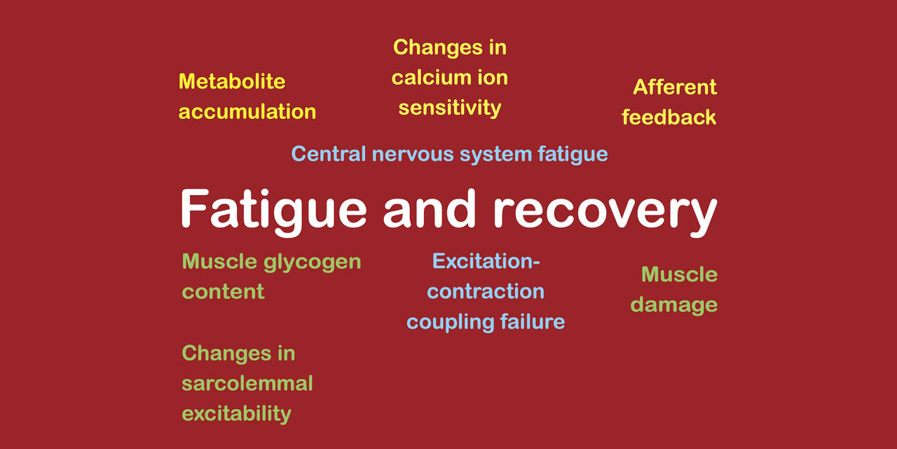
When is each fatigue mechanism present?
Please subscribe to my newsletter to read the future articles in this series!
In the fitness industry (as well as in real life), fatigue is often referred to as a single phenomenon that builds up during exercise, reaches a peak at the end of exercise, and then dissipates afterwards. This is completely false. Fatigue is made up of different mechanisms, some of which are apparent immediately upon commencing exercise, others do develop slowly during exercise, and yet others are not present during exercise and in fact only appear hours after the bout of exercise has finished. In fact, exactly when each fatigue mechanism is present is a challenge to identify!
In my first article in this series, I explained how fatigue can be analyzed by looking at its four main outputs (reduced coordination, reduced motor unit recruitment, reduced muscle fiber shortening velocity, and reduced muscle fiber force). My second article provided further detail about the locations of each of the fatigue mechanisms that generate each of those outputs. In this third article, I aim to show that different fatigue mechanisms develop at different rates and are therefore present at different times, which is another reason why it helps to understand the variety of fatigue mechanisms that exist, rather than treating fatigue as a single, unified construct.
When exactly is each fatigue mechanism present?
#1. Impairment in the generation of central motor command, which is known as supraspinal central nervous system (CNS) fatigue
Under certain circumstances, the level of central motor command (CMD) that the brain is able to produce is reduced, despite us attempting to exert the greatest possible effort. This leads to a reduction in the level of motor unit recruitment in the working muscle, which in turn reduces muscle force, which we then measure as fatigue. Since it is motor unit recruitment that is affected and not muscle fiber force or shortening velocity, we refer to this type of fatigue as CNS fatigue. And since the mechanisms are present inside the brain, we refer to the CNS fatigue as supraspinal CNS fatigue, so as to differentiate it from spinal CNS fatigue.
Supraspinal CNS fatigue occurs when the CMD produced by the brain is lower than expected, despite us exerting a maximal effort. The limiting factor for the brain producing a CMD is our maximum tolerable level of perceived effort, which itself can be modified slightly from day to day by our motivation levels. In other words, the amount of CMD that the brain is able to produce depends on [1] the perceived level of effort that is experienced at any given level of motor unit recruitment, and [2] the level of motivation that is experienced. By definition, supraspinal CNS fatigue must therefore occur when either [1] the perceived level of effort experienced at a given level of motor unit recruitment is increased, or [2] when motivation is reduced.
This is important, because it means that the rate at which supraspinal CNS fatigue develops can vary according to the source of the factor that increases the perception of effort or which reduces the level of motivation!
Some factors appear immediately upon commencing exercise. For example, we could argue that the suppressed central motor command during eccentric contractions (caused by the additional complexity of the contraction mode leading to an increased perception of effort) is in fact a form of supraspinal CNS fatigue that appears even before the production of muscular force. In the same way, when environmental factors are in place that inhibit motivation, supraspinal CNS fatigue is created even before muscle force is generated.
Other factors develop during exercise. For example, in a strength training set, metabolite accumulation occurs that produces afferent feedback to the brain that we experience as fatiguing and burning sensations inside the muscle. This increases the perception of effort for the existing level of motor unit recruitment, thereby producing supraspinal CNS fatigue. Similarly, during aerobic exercise, inflammatory mediators are slowly released into the bloodstream that also produce afferent feedback to the brain that we probably experience as more systemic fatigue sensations. In the same way, this increases the perception of effort for the existing level of motor unit recruitment, thereby producing supraspinal CNS fatigue.
And other factors only develop after stopping exercise. After an eccentric training workout, muscle damage occurs that stimulates an inflammatory response several hours after exercise. The local and systemic inflammatory responses then create afferent feedback to the brain that we probably experience as systemic fatigue sensations. Again, this increases the perception of effort for the existing level of motor unit recruitment, thereby producing supraspinal CNS fatigue.
In summary, when supraspinal CNS fatigue occurs can vary to an incredible extent, depending on what underlying mechanism is involved in causing a change in either the perception of effort or the level of motivation. Some factors cause supraspinal CNS fatigue either before or upon starting exercise, other factors cause supraspinal CNS fatigue to develop during exercise (and at varying rates), and other factors cause supraspinal CNS fatigue to develop only hours after exercise. Therefore, whether supraspinal CNS fatigue is present at any moment in time (during or after exercise) depends greatly upon how it was produced in the first place.
#2. Impaired transmission of central motor command, which is known as spinal CNS fatigue
Under certain circumstances, the level of CMD that the spinal cord is able to transmit is reduced, despite the brain producing a high CMD. This leads to a reduction in the level of motor unit recruitment that reaches the working muscle, which in turn reduces muscle force, which we then measure as fatigue. Since it is motor unit recruitment that is affected and not muscle fiber force or shortening velocity, we refer to this type of fatigue as CNS fatigue. And since the mechanisms work inside the spinal cord, we refer to the CNS fatigue as spinal CNS fatigue, to differentiate it from supraspinal CNS fatigue.
Perhaps more than any other fatigue mechanism, spinal CNS fatigue during exercise works much like the basic fatigue models predict. Spinal CNS fatigue seems to be dependent upon the duration of time for which a motor unit is firing. Thus, the longer the motor unit fires, the more spinal CNS fatigue develops. Even so, it seems likely that spinal CNS fatigue dissipates incredibly quickly after cessation of exercise, perhaps even within minutes. Thus, the rate of development is probably quite a bit slower than the dissipation rate.
#3. Reduced propagation of electrical signal within muscle, which is often described as a reduction in sarcolemmal excitability
For muscle fibers to exert force, they must be activated. This activation occurs by means of the propagation of electrical signals along the cell membrane or sarcolemma of the muscle fibers themselves. Yet, when the polarity of the cell membrane is disrupted, the propagation of the electrical signal is interrupted, which prevents the activation of the muscle fiber. When this happens, some of the muscle fibers in the muscle, which were signaled to switch on and exert force, fail to activate. This reduces muscle force, which we measure as fatigue.
This impairment in the ability of electrical signals to propagate along the cell membrane may occur at different times or rates, depending on the underlying mechanism that is responsible. Since the polarity of the cell membrane is determined by the presence of ions on either side, ionic shifts during exercise can cause changes in the ability of electrical signals to propagate. Such shifts can occur relatively quickly during exercise, but likely also dissipate quickly after cessation of exercise. Also, since the polarity of the cell membrane rests upon the integrity of the cell membrane, any damage to the membrane can cause changes in the ability of electrical signals to propagate. Cell membrane damage is caused by reactive oxygen species (ROS) that are released during exercise, which trigger the release of phospholipases that can degrade the membrane. This multi-stage process likely takes longer to occur, and the involvement of structural damage implies that it will also take longer to dissipate once it has occurred. Indeed, when meaningful reductions in sarcolemmal excitability have been observed, they have been recorded as occurring 15 minutes after finishing exercise and lasting for several hours after exercise. The slight delay likely reflects the time required for the ROS to trigger the release of phospholipases, and for the phospholipases to damage the cell membrane. The several hours for dissipation to occur likely reflects the repair process of the cell membrane.
#4. Impaired conversion of an electrical signal into chemical signal, which we call “excitation-contraction coupling failure” (ECCF)
For muscle fibers to produce force, their myofibrils must detect a chemical signal that takes the form of calcium ions released from stores within the sarcoplasmic reticulum. For the chemical signal to be generated, the electrical signal that was propagated along the outside of the muscle fiber cell membrane and down into the transverse tubule must be converted into the chemical signal at the triadic junction with the sarcoplasmic reticulum. This conversion process is called excitation-contraction coupling. When a fatigue mechanism happens that stops the electrical signal triggering the release of calcium ions at the triadic junction, the myofibrils inside the muscle fiber do not receive a chemical signal that tells them to form crossbridges, so they do not make any. When this happens, some of the muscle fibers, which were fully activated, do not produce force in spite of being activated. We call this ECCF. This in turn reduces muscle force, which we measure as fatigue.
ECCF is produced when too many calcium ions fail to return to the sarcoplasmic reticulum after being placed there during the excitation-contraction coupling process. Their presence stimulates the release of calpains, which then degrade the minor proteins that hold the triadic junction in position, which allows the voltage sensor that detects the arrival of the electrical signal to drift away from the sarcoplasmic reticulum calcium ion store, thereby preventing the electrical signal from triggering the release of calcium ions into the cytoplasm. Since this process involves multiple phases, including the accumulation of calcium ions inside the muscle fiber, the release of calpains, and the damage occurring at the triadic junction, it takes time to develop during exercise, and even longer to dissipate afterwards.
#5. Impairment in the detection of chemical signal by actin myofibrils, leading to a loss of calcium ion sensitivity
For muscle fibers to produce force, their myofibrils must detect a chemical signal that takes the form of calcium ions released from stores within the sarcoplasmic reticulum. Yet, various events can occur within the muscle fiber cytoplasm that stop actin myofibrils from detecting the presence of calcium ions. Therefore, even when calcium ions are being adequately supplied by the sarcoplasmic reticulum, the actin myofibrils can still fail to form crossbridges, simply because they cannot detect the calcium ions. When this happens, some of the muscle fibers, which were fully activated, do not produce force in spite of being activated. This reduces muscle force, which we measure as fatigue.
Exactly how quickly calcium ion sensitivity is lost during exercise, as well as how long the fatigue mechanism takes to dissipate afterwards, are relatively unclear (because the exact mechanisms that cause each to occur are also fairly unclear), but the loss of calcium ion sensitivity probably occurs faster than ECCF and also dissipates much more rapidly.
#6. Slowing or preventing of the binding of myosin heads to actin myofibrils
For muscle fibers to produce force, their myofibrils must form crossbridges, and the rate of crossbridge binding and unbinding is what determines muscle fiber force and muscle fiber shortening velocity. Therefore, any fatigue mechanism that causes slowing of either binding or unbinding can affect muscle fiber force or muscle fiber shortening velocity.
Indeed, both acidosis and the production of inorganic phosphate as a result of the use of ATP slow or even stop the rates of crossbridge binding and unbinding. Importantly, acidosis seems to affect muscle fiber shortening velocity more than force (and generates its effects more quickly), while phosphate seems to affect muscle fiber force more than shortening velocity (and produces its effects more slowly).
Thus, acidosis occurs fairly quickly during a fatiguing muscular contraction (leading to early reductions in muscle fiber shortening velocity), while the accumulation of phosphates occurs later on (leading to later reductions in muscle fiber force). This discrepancy has important implications for strength training for strength, hypertrophy, and speed, since strength and hypertrophy are developed by high levels of muscle fiber force, while speed is increased by high levels of muscle fiber shortening velocity. Both metabolites dissipate quickly after muscular contractions stop, such that any fatigue that they cause is gone within 30 minutes after exercise.
#7. Transmission of crossbridge force to the surrounding cytoskeleton
For muscles to produce force, the shortening of myofibrils inside muscle fibers by the formation of actin-myosin crossbridges must lead to the shortening of the muscle fiber itself. This occurs by the transmission of forces from the myofibrils to the cytoskeleton and through the cell membrane to the surrounding endomysium of the muscle fiber. Yet, if any of the myofibrillar or cytoskeletal structures are damaged, then this transmission of crossbridge forces is impaired, even though the muscle fiber might be fully activated, even though calcium ions might be flowing freely into the cytoplasm, and even though many of the actin myofibrils inside the muscle fiber might be responding to the presence of those calcium ions to form crossbridges. This reduction in muscle fiber force in turn reduces whole muscle force, and is then detected as fatigue.
Importantly, while it is often assumed that the mechanical tension generated by muscle fibers during muscular contractions is what causes muscle damage to occur, this is not actually the case. In fact, muscle damage is caused by the accumulation of calcium ions inside the cytoplasm leading to the stimulation of calpains that degrade the cytoskeleton and the myofibrils. In this way, muscle damage follows the same process as ECCF, except that ECCF involves damage to very specific, very small proteins associated with the triadic junction, while muscle damage involves more generalized damage. Also, muscle damage seems to take longer to become apparent (indeed, it is not apparent during or immediately after exercise, but only appears after several hours), perhaps because the actions of calpains take longer to affect the larger structures inside the muscle fibers and because the structures are more widely spread throughout the cytoplasm. Thus, muscle damage is one of the few types of fatigue that is actually not at all present during exercise, and in fact only appears after exercise has ceased.
Why is this important? (in theory)
As explained in the above analysis, fatigue is made up of different mechanisms that are present at different points in time. Some are apparent even before starting exercise (as when motivation is negatively affected), some of which are caused upon commencing exercise (as when a complex movement is performed that requires a great deal of cognitive processing), some of which occur during exercise and then dissipate quite soon afterwards (as when metabolites build up), some of which occur during exercise and then dissipate slowly afterwards (as when calcium ions cause ECCF), some of which only become apparent just after exercise (as when ROS cause phospholipases to digest the cell membrane and impair electrical signal propagation), and some of which only become apparent hours after exercise (as when calcium ions cause muscle damage and the resulting inflammatory response causes supraspinal CNS fatigue).
Importantly, these fatigue mechanisms can impair either [A] coordination, or [B] motor unit recruitment level, or [C] maximum muscle fiber force, or [D] maximum muscle fiber shortening velocity. When one of these outputs is impaired during exercise, the stimulus for it to improve after exercise is reduced. However, every fatigue mechanism does not impair all of these outputs to the same extent. As explained in the above analysis, some of them only affect motor unit recruitment level, while others only affect either muscle fiber force or muscle fiber shortening velocity. Thus, the presence of a certain mechanism at a certain point in time is critical for understanding whether an adaptation can be stimulated, despite some fatigue being present.
Why is this important? (an example)
Supraspinal CNS fatigue can reduce the magnitude of the CMD during a bout of exercise performed when it is present. By doing this, it prevents gains in the ability to recruit high-threshold motor units (HTMUs) from occurring, which is an essential mechanism through which gains in maximum strength develop. Fortunately, by looking at the various ways in which supraspinal CNS fatigue occurs, we can see how to structure training programs that so that we do not bother trying to do workouts that aim to increase the ability to recruit HTMUs when supraspinal CNS fatigue is present.
Supraspinal CNS fatigue is present when motivation is reduced – Therefore, there is little point in attempting to stimulate an improvement in the ability to recruit HTMUs when an athlete is demotivated due to factors related to the training program (such as being at the end of a high volume training block) or to the sport (such as performing in a congested schedule).
Supraspinal CNS fatigue is present when complex movements are performed – because these involve additional cognitive processing, which increases the perception of effort. Therefore, there is little value in attempting to to stimulate an improvement in the ability to recruit HTMUs in novel exercises or in complex exercises that the athlete is not very experienced in performing.
Supraspinal CNS fatigue is present when metabolite accumulation occurs (because the metaboreceptors inside the muscle detect the presence of lactate, acidosis and inorganic phosphate and send afferent feedback to the brain). Therefore, it is unlikely that light load strength training to failure will be able to produce meaningful improvements in the ability to recruit HTMUs (conversely, cluster sets and velocity-based training methods may be particularly effective).
Supraspinal CNS fatigue is present when inflammatory mediators build up over the course of a workout (due to afferent feedback and also due to the detection of the mediators in the bloodstream. Thus, is unlikely that sets or exercises performed at the end of a workout will be able to produce meaningful improvements in the ability to recruit HTMUs.
Supraspinal CNS fatigue is present when inflammatory mediators build up after a workout due to muscle damage. Thus, is unlikely that workouts that are performed while substantial muscle damage is still present from a previous workout will be able to produce meaningful improvements in the ability to recruit HTMUs.
As this example demonstrates, there are very specific situations in which the presence of supraspinal CNS fatigue can stop a certain, important adaptation from occurring. Knowledge of why this happens is valuable, because it enables us to structure workouts to focus our efforts on stimulating the adaptation when it is actually possible to stimulate the adaptation. Nevertheless, this is only one example. The same information can be gathered for other types of fatigue that affect muscle fiber force or muscle fiber shortening velocity.
What is the takeaway?
Characterizing fatigue as a single phenomenon is an oversimplification that makes it difficult to see how it develops during exercise and then dissipates afterwards. In contrast, if we break fatigue down into various mechanisms, we can see when each type of fatigue develops and then dissipates through each of these mechanisms. Since each type of fatigue impairs the development of different adaptations (such as improved coordination, improved ability to recruit HTMUs, improved muscle fiber size and force, and improved muscle fiber shortening velocity), we can then avoid trying to develop each of these qualities when the wrong type of fatigue is present. For example, we can avoid trying to improve the ability to recruit HTMUs when CNS fatigue is present (because the CNS fatigue will stop it happening). But also, we can avoid trying to improve muscle fiber size and force when muscle fiber force is impaired (because the reduced mechanical tension stops these adaptations occurring), and we can avoid trying to improve speed when muscle fiber shortening velocity is temporarily reduced. For this reason, knowing exactly when each type of fatigue mechanism is present (both during a workout and also in the post-workout recovery period) is very important.